علت چروکیدگی انگشتان بعد از حمام!

حتما بعد از حمام دیده‌اید که دست‌هایتان مانند آلوی خشک چروکیده شده است. دانشمندان بر خلاف باور عامه این پدیده را ناشی از فشار آب بر پوست نمی‌دانند؛ بلکه دریافته‌اند که این امر ناشی از تکامل سیستم عصبی انسان است تا چسبندگی دست به اشیاء در حالت مرطوب تقویت شود.

چرا پوست انگشتان انسان پس از خیس خوردن در حمام مانند آلوی خشک چروک می‌خورد؟ دانشمندان فکر می‌کنند به این سوال پاسخ داده‌اند. آزمایش‌ها این نظریه را تایید نموده‌اند که انگشتان چروکیده، چسبندگی دست ما را با اشیاء خیس بهتر می‌کنند؛ مانند آبی که روی عاج لاستیک‌های ماشین جاری باشد. اغلب افراد چروکیدگی و تورم پوست را نتیجه‌ فشار آب بر لایه بیرونی پوست می‌دانند. اما پژوهشگران از دهه ۱۹۳۰ می‌دانند وقتی آسیب عصبی در انگشتان وجود داشته باشد، این اثر رخ نمی‌دهد، که نشانگر تغییر در اثر واکنش غیرارادی توسط سیستم عصبی خودمختار بدن است – سیستمی که تنفس، ضربان قلب و تعریق را کنترل می‌کند. در واقع، چروکیدگی دیگری وجود دارد که ناشی از انقباض رگ‌های خونی زیر پوست است.

در ۲۰۱۱، مارک چنگیزی (Mark Changizi) یک عصب-زیست‌شناس تکاملی در آزمایشگاه‌های 2AI بویزی، آیداهو در ایالات متحده و همکارانش پیشنهاد کردند که چروکیدگی – که فرایندی فعال است – باید عملکردی تکاملی داشته باشد. همچنین نشان دادند که الگوی چروکیدگی برای تهیه شبکه زهکشی و افزایش چسبندگی بهینه به‌نظر می‌رسد. اما تاکنون هیچ اثباتی وجود نداشت که نشان دهد انگشتان چروکیده در واقع یک مزیت هستند. در آخرین مطالعه، شرکت‌کنندگان اشیاء تر یا خشکی مانند تیله را با اندازه‌های مختلف با دست معمولی یا انگشتان متورم بعد از غوطه‌ورسازی در آب داغ به مدت ۳۰ دقیقه، می‌گرفتند. افراد در گرفتن تیله‌های تر (نسبت به تیله‌های خشک) با دستان چروکیده موفق‌تر بودند اما چروک‌ها تفاوتی در حمل اشیای خشک ایجاد نمی‌کردند؛ نتایج در مجله بایولوژی لترز چاپ شده‌اند.

تام اسمولدرز (Tom Smulders)، زیست‌شناس تکاملی در دانشگاه نیوکاسل انگلستان و نویسنده همکار در این مقاله می‌گوید: «ما نشان داده‌ایم که انگشتان چروکیده در شرایط مرطوب چسبدگی بهتری دارند. در واقع همانند عاج چرخ‌های خودروی شما عمل می‌کنند که باعث تماس بیشتر لاستیک با جاده می‌شوند.» اسمولدرز می‌افزاید: «شاید انگشتان چروکیده به اجداد ما کمک کرده‌اند تا غذا را از پوشش‌های گیاهی یا جریان های مرطوب بگیرند.» اثر مشابهی در انگشتان پا کمک می‌کند تا در باران راحت‌تر گام برداریم.

چنگیزی می‌گوید این نتایج مدرکی رفتاری از این هستند که «انگشتان شبیه آلوی خشک، مانند عاج لاستیک در برابر باران عمل می‌کنند» که با یافته‌های ریخت‌شناسی گروه او سازگار است. او می‌افزاید باید بررسی کنیم که آیا چروکیدگی‌ مشابه با همین مزیت در دیگر حیوانات رخ می‌دهد یا خیر. «هنوز نمی‌دانیم که چه گونه‌هایی جز انسان و بوزینه این ویژگی را دارند.» چروک‌ها در رابطه با اشیاء تر مزیتی را به ما اعطا می‌کنند، اما به‌ظاهر با اشیاء خشک مشکلی ندارند. اسمولدز می‌گوید مشخص نیست که چرا انگشتان ما به شکل دائمی چروکیده نیستند؛ اما او ایده‌هایی دارد: «برداشت اولیه ما می‌گوید شاید این موضوع حساسیت سرانگشت‌های ما را از بین ببرد یا خطر آسیب از طریق گرفتن اجسام را افزایش دهد.»

Proteins boost quantum coherence in bacteria

A new theory of how plant photosynthesis involves quantum coherence has been suggested by physicists in the UK, Germany and Spain. This latest research is based on the study of organisms that live deep under the sea yet are able to convert sunlight into energy. The study suggests that molecular vibrations do not destroy the coherence – as previously thought – but rather perpetuate and even regenerate coherence. The discovery provides a better understanding of how as much as 99% of the energy of light absorbed by photosynthesis cells is successfully transferred to locations in the cells where electric energy is converted to chemical energy. The work opens up the possibility of using nature-inspired designs in quantum devices.

Until recently, living systems were thought to be "too wet and warm" to rely on delicate quantum properties such as entanglement and coherence. The problem is that these properties decay rapidly via random interactions with things in the outside world, such as vibrating molecules. However, over the past decade physicists have begun to suspect that quantum properties play important roles in biochemical processes – including photosynthesis. This latest work was done by Alex Chin (now at Cambridge University) and colleagues at the Institute of Theoretical Physics in Ulm and the Technical University of Cartagena. The team looked at organisms called green sulphur bacteria that live 2000 m below the ocean surface. There is so little sunlight down there that the bacteria cannot afford to lose a single photon – indeed, almost 100% of the light they absorb is turned into food.

Excited states

When sunlight hits the surface of the plant, energy is transferred via chains of pigments to a reaction centre, where it is converted into chemical energy. Those pigments are held in place by proteins, which together create pigment–protein complexes, or PPCs. The PPCs effectively act as corridors and the energy itself travels in the form of molecular excited states, or molecular excitons. These excitons are able to move along the PPC by hopping from one molecule to the next.

In 2007 Graham Fleming and colleagues in the US showed that these excitons exhibit quantum coherence, which means that the excitons may exist simultaneously in a superposition of several quantum states with varying probabilities. Coherence also allows the exciton to explore multiple pathways to the reaction centre simultaneously, ultimately choosing the fastest, most efficient option. As is demonstrated in man-made solar cells (which also rely on excitons), the longer this trip takes, the more likely it is that the energy will dissipate before it reaches its destination.

Optimizing function

The presence of quantum effects in photosynthesis surprised both physicists and biologists, and left them wondering how a fragile quantum state could survive in a living organism. More specifically, research groups found that the coherenent states exist for 100-times longer than the coherence time of the energy states of an exciton. Something was helping these wave states survive long enough to ensure the safe passage of nearly 100% of the photon energy that the organisms absorbed.

This latest research suggests that the answer lies in the proteins in the PPCs, which provide structural support for the pigment molecules. The new calculations reveal that these proteins are more active participants in the transport system than was previously thought. The natural vibration frequencies of the proteins resonate with the exciton waves, and like a parent pushing a child on a swing, the protein structures keep the excitons oscillating without dampening. In fact, the exciton may pass its vibration into the protein structures, which then return it to the exciton, thus restoring its coherence. "People have not viewed this protein structure as something that actively helps quantum phenomenon to take place in biological systems," says team member Martin Plenio. "This is really a new way of thinking about things."

Definitely not noise

The team's conclusions come from precise analysis of the protein vibrations, using data from Markus Wendling and colleagues in the Netherlands, who in 2000 examined the PPC structures from green sulphur bacteria. Previous efforts to study the protein vibrations used rougher approximations and usually concluded that the vibrations were noise.

"The main difference in terms of the paradigm for doing this simulation was to not separate the system into the exciton and the environment, but to treat them all together as one large many-body system," says Chin. "We took a completely holistic approach. This makes it very complicated in terms of variables and things that one has to keep track of, which means that computationally it is very tough."

Understanding these protein structures could assist in building similar structures in quantum devices. If similar structures are used in the conversion of electrical energy to chemical energy, it could shed light on how to mimic photosynthesis's high efficiency rates in man-made solar cells.

A good hypothesis

Greg Scholes of the University of Toronto in Canada praises the detailed analysis conducted by the team, and says the conclusions "fit with some of the pieces of the puzzle that have been emerging in more recent experiments".  While Scholes believes the analysis is "sufficient proof of the idea in principle", he says direct experiments will need to be done to confirm the conclusions. "From that perspective this work really contributes something important. Because it gives us a hypothesis, [and now] we can go and test it," he says.

طبقه‌بندی وول خوردن‌های پروتئینی

تغییر شکل پروتئین‌ها که برای پیش‌برد فرآیندهای شیمیایی ضروری است به دو شکل ارتعاشات کشسان و تغییرات صورتبندی صورت می گیرد. تشخیص این دو نوع تغییر اهمیت ویژه‌ای در درک عملکرد پروتئین‌ها در بدن موجودات زنده ایفا می‌کند. محققان آزمایشگاه ملی اوک ریج آمریکا به تازگی روش جدیدی را برای تشخیص ارتعاشات کشسان و تغییرات صورتبندی از یکدیگر معرفی کرده‌اند.

پروتئین‌ها برای این که به درستی کار کنند، به تغییرات شکل‌شان متکی هستند. بنابراین دانشمندان به تغییرات کوچک در ساختار پروتئین‌ها توجهی جدی دارند، هر چند که تمام این «تکان‌ها» مانند هم نیستند. مطالعه جدیدی که آزمایش‌های پراکندگی نوترون و نور را با شبیه‌سازی دینامیک مولکولی تلفیق کرده است، نشان می‌دهد که چگونه می‌توان با استفاده از اندازه‌گیری انعطاف‌پذیری پروتئین‌ها، تفاوت میان ارتعاشات کشسان و تغییرات صورتبندی (conformational) واقعی را تشخیص داد. نتایج این مطالعه که در فیزیکال ریویو لترز ارائه شده است، رفتارهای خاصی از پروتئین‌ها را توضیح می‌دهد، از جمله اینکه چرا انعطاف‌پذیری پروتئین‌ها گاهی به سرعت با دما افزایش می‌یابد.


برای پیش‌برد فرآیندهای شیمیایی، پروتئین‌ها اغلب مجبورند که به شکل فیزیکی (مثل کلید) با دیگر مولکول‌ها چفت شوند. پروتئين‌ها می‌توانند با وول خوردن و لرزیدن سر جای خودشان، دچار تغییرات صورتبندی شوند که در آن اتم‌های درون مولکول از یک سد انرژی پتانسیل به سد انرژی دیگر می‌جهند. از طرف دیگر، انعطاف‌پذیری پروتئین‌ها نیز از اتم‌هایی ناشی می‌شود که به شکل کشسان درون یک سد پتانسیل افت‌ و‌ خیز می‌کنند. تغییرات کشسان و صورتبندی باید به شکل متفاوتی به دما و آب‌پوشی مولکول وابسته باشند، اما مطالعات قبلی درباره انعطاف‌پذیری پروتئین‌ها معمولا این دو سهم را جدا نکرده‌اند.

به تازگی لیانگ هونگ (Liang Hong) از آزمایشگاه ملی اوک ریج آمریکا و همکارانش راهی موثر برای جداسازی حرکت‌های کشسان و صورتبندی از یکدیگر معرفی نموده‌اند. آن‌ها در ابتدا انعطاف پذیری را – همان‌طور که اغلب انجام می‌شود – با استفاده از پراکندگی نوترون‌های ناهمدوس اندازه‌گیری نموده و جابجایی‌های اتمی (عمدتا اتم‌های هیدروژن) درون یک پروتئين خاص را ثبت کردند. سپس با مقایسه این داده‌های جابجایی با شبیه‌سازی دینامیک مولکولی گروه هونگ دریافتند که افت و خیزهای کشسان اتم‌های منفرد را می‌توان از ارتعاشات نوسانی کل مولکول که آن‌ را با آزمایش‌های پراکندگی نور اندازه‌گیری کرده بودند استنتاج نمود. این پژوهشگران با جداسازی بخش کشسان در واقع نشان دادند که انعطاف‌پذیری بالا در دمای اتاق اصولا ناشی از آهنگ بیشتر تغییرات صورتبندی آن است.

لینک منبع

تاخوردگی سریع DNA: راهی برای ساخت ماشین‌های زیستی نانومقیاس

مطالعه تازه‌ای نشان می‌دهد که رشته‌های DNA را می‌توان طی چند دقیقه به اشکال گوناگون در‌آورد. این یافته‌ها می‌تواند سرعت پیشرفت حوزه اوریگامی DNA را شدیدا افزایش دهد و تحولی در ساخت ماشین‌های نانومقیاس ایجاد نماید.

بیوتکنولوژی‌دانان قصد دارند تا از DNA، ماشین‌های نانومقیاس درست کنند اما تاکنون تنها اشکال ساده‌ای مانند لوله، جعبه یا مثلث ساخته‌اند زیرا این فرایندها طاقت‌فرسا و زمان‌بر هستند. این روش‌ها از رشته‌های DNA برای نگهداری رشته‌های تاشده و طولانی‌تر در نقاط خاصی مانند نوارچسب استفاده می‌کنند. تاکنون، شکل‌دهی را با گرم‌سازی DNA و سپس سردسازی آهسته آن تا یک هفته انجام می‌شد.

اما اکنون این زمان به دقیقه تخفیف یافته است. هندریک دیتز (Hendrik Dietz) بیوفیزیکدانی در دانشگاه فنی مونیخ و همکارانش، DNA را با رنگ فلورسنت رنگ کردند و آن چه را در هنگام سردسازی و تا زدن رخ داد، تماشا کردند. با توقف واکنش در سطوح مختلف، آن‌ها می‌توانستند ببینند که تا خوردن و چسبیدن تا چه حدی پیش رفته است.

آن‌ها چیز عجیبی کشف کردند؛ دیتز می‌گوید: «معلوم شد که تقریبا در تمام بازه دمایی هیچ اتفاقی نمی‌افتد.» اما وقتی به دمای خاصی می‌رسیم، ناگهان کل ساختار شکل می‌گیرد. این نتایج در ساینس منتشر شده‌اند. دیتز تا خوردن ۱۹ شکل گوناگون DNA مانند اشیا استوانه‌ای، آجرمانند و  چرخ‌دنده مانند را تحلیل کرد. تمام اشکال در بازه دمایی کوچکی بین ۴۵ تا ۶۰ درجه سانتی‌گراد تا می‌خوردند.

 اشکال پیچیده‌تر

بعد از استخراج این‌که کدام دما با کدام شکل متناظر است، دیتز مخلوط‌های واکنشی DNA تانخورده را برای مدت چند دقیقه تحت این دماهای از پیش تعیین شده گذارد تا ببیند آیا به شکل مطلوب تا می‌خورند؛ با بازدهی بالایی همین‌طور بود.

متخصصان اوریگامی DNA از شتاب پیشرفت کارهای خود هیجان‌زده شده‌اند. ویلیام شیه (William Shih) که در دانشگاه هاروارد کار می‌کند و گروهش از کار دیتز منتفع شده‌ است، می‌گوید: «این کار زندگی ما را اندکی ساده‌تر می‌کند.» تا زدن آسان‌تر، سریع‌تر و کاراتر به اوریگامی DNA کمک می‌کند تا به ورای شکل‌های ساده‌ رود.

دیتز امید دارد که بتواند یافته‌های خود را برای ساخت مدلی رایانه‌ای استفاده کند که چگونگی ساخت دیگر اشیا DNA را پیش‌بینی نماید. او می‌گوید که خصلت اشیا با دمای تا خوردنشان همبستگی دارد – مثلا اشیایی که از رشته‌های طولانی‌تر ساخته‌ شده‌اند، در دمای بالاتری تا می‌خورند. دیتز قصد دارد تا نانوساختارهایی با دماهای تاخوردن بهینه نزدیک به ۳۷ درجه سانتی‌گراد بسازد؛ دمایی که سلول پستانداران در آن رشد می‌کند. به این ترتیب شاید بتوان روزی از ماشین‌های DNA در شرایط زیستی استفاده کرد.

لینک منبع مقاله

Complex 3D nanostructures built using DNA bricks

A new technique to make highly complex 3D nanostructures by assembling together synthetic DNA "bricks" has been developed by researchers at Harvard University in the US. The bricks, which are like tiny pieces of LEGO, can be assembled into a wide variety of shapes and configurations, meaning that they can be used to build elaborately designed nanostructures. The resulting structures might find use in a wide variety of applications, including smart medical devices for targeted drug delivery in the body, programmable imaging probes and even in the manufacture of speedier and more powerful computer-chip circuits. DNA nanotechnology has now been around for nearly 30 years, but it really took off with the advent of a technique called DNA "origami". This technique, named after the ancient Japanese art of paper folding and first developed in 2006 by Paul Rothemund at the California Institute of Technology, involves folding long strands of DNA into a wide range of predetermined shapes. The resulting nanostructures can be used as scaffolding or as miniature circuit boards for precisely assembling components such as carbon nanotubes and nanowires.

Powerful though it is for making both 2D and 3D shapes, DNA origami has its limitations. To fold the DNA, several hundred "staples" must be added to the regions surrounding the single DNA strands, and each type of new nanostructure desired requires a new set of staples. Moreover, the DNA structures tend to arrange themselves randomly onto a substrate surface, which makes it difficult to integrate them into electronic circuits afterwards.

Building bricks

A team led by Peng Yin at Harvard first put forward its DNA-brick self-assembly technique earlier this year. Rather than starting with long DNA strands, the researchers succeeded in interlocking short, synthetic strands of DNA together to make larger structures. In fact, they managed to arrange the short strands into a "molecular canvas" by controlling the local interactions between the strands. The technique, like any DNA self-assembly method, works by exploiting the fact that the four base pairs in DNA – adenosine, thymine, cytosine and guanine – are naturally programmed to join up in specific ways: A only binds to T, while C only binds to G. So, the team was able to fabricate a collection of 2D structures using its technique by stacking one DNA brick that was 42 bases long upon another brick.

3D shapes

Now, Yin and colleagues have extended their technique to 3D. The researchers begin with an even smaller DNA-brick strand – only 32 bases long – that contains four regions than can bind to four neighbouring DNA-brick strands. The bricks are connected through 90° and so can be built out in all three directions – up, down, and out – to create a solid "master" DNA molecular-canvas cube containing hundreds of bricks. Compared with hand-assembled LEGO structures, each DNA structure self-assembles thanks to the fact that every brick is encoded with an individual sequence that determines its final position in the nanostructure. Each sequence will only be attracted to one other complementary sequence, which means that specific shapes can be created through the selection of different sequences.

The biggest advantage of the new DNA-brick technique is that any number of structures can effortlessly be made from the same master cube by simply selecting subsets of specific DNA bricks, according to the team. "We have already made more than 100 different shapes in this way (with some containing intricate cavities, surface features and channels), all of which are more complex than any 3D DNA structure constructed in the last decade. What is more, additional DNA bricks can be added, removed or modified independently without affecting other parts of the structure," says Yin.

Complex structures

The researchers claim that the complex structures that can be made using their DNA-brick assembly technique will help advance existing DNA nanotechnology applications. "We can for example, arrange technologically relevant guest molecules into functional devices that might serve as programmable molecular probes, instruments for biological imaging and drug-delivery vehicles," Yin tells. "The structures can also be used to fabricate high-throughput complex inorganic devices for electronics and photonics applications."

The DNA-brick structures are also entirely synthetic, whereas DNA origami is half biological. This expands the range of potential applications even further, Yin adds. "For instance, by using synthetic polymers rather than the natural form of DNA, we might be able to create functional structures that are stable in a wider variety of different environments." The team is now busy improving its brick technique by looking more closely at DNA structure and sequence design, enzymatic synthesis for higher-quality strands and optimizing processing conditions. "We would also like to better understand the kinetic pathways involved in DNA assembly," says Yin.

باکتری‌ها الکترون‌ها را در فواصل سانتی‌متری هدایت می‌کنند.

به تازگی گونه‌ای باکتری در اعماق دریا کشف شده است که الکترون‌ها را در طی فواصل سانتی‌متری هدایت می‌کند تا بتواند در محیطی با اکسیژن کم، با استفاده از سولفید هیدروژن تغذیه کند. این ادعای دانشمندانی در دانمارک و آمریکاست. آن‌ها نشان داده اند که هزاران میکرو ارگانیسم از یک رشته که یک سر آن‌ها به رسوبات اقیانوس و سر دیگرشان به آب شور می‌رسد، این کار را انجام می‌دهند. فهم این موضوع که این موجود زنده چگونه الکترون‌ها را انتقال می‌دهد، می‌تواند منجر به تکنولوژی جایگزینی برای تولید انرژی شود.


زیست شناسان می‌دانستند که خانواده دی‌سولفو‌بولباکی از باکتری‌ها با مصرف ترکیبات سولفور در اقیانوس خود را تقویت می‌کنند. این موجب تولید سولفید هیدروژن می‌شود که در غلظت‌های بالا سمی است. زمانی‌که این باکتری سولفید هیدروژن مصرف می‌کند باید در حضور اکسیژن باشد که طی این واکنش انتقال الکترون صورت گیرد. رسوبات اقیانوس معمولاً سطح اکسیژن خیلی پایینی دارند و مطالعات نشان داده است زمانی که دی‌سولفو‌بولباکی وجود دارد، سطح سولفید به طور یکنواخت و پیوسته افزایش می‌یابد. اما سپس اتفاق پیش‌بینی نشده‌ای می‌افتد. سطح سولفید به سرعت افت پیدا می‌کند انگار که رسوبات یک هجوم ناگهانی از اکسیژن را تجربه کرده‌اند. مسئله اینجاست که این افت به قدری سریع است که با نظريه‌ی پخش مولکول‌های هیدروژن قابل توجیه نیست. در عوض دانشمندان اندیشیده‌اند که نمونه‌های متفاوت بسیاری از این نوع باکتری در رسوبات  وجود دارند و به طریقی الکترون‌ها را از نواحی با اکسیژن پایین به سمت آب شور که میزان اکسیژن موجود در آن زیاد است انتقال می‌دهند.

باکتری‌های منفرد

اکنون تیمی از فیزیک‌دانان دانشگاه کالیفرنیای جنوبی و دانشگاه آرهیوز از جمله محمد الناگر نگاه دقیق‌تری به باکتری‌های موجود در رسوبات داشته‌اند و کشف شگفت‌انگیزی کرده‌اند مبنی بر اینکه انتقال الکترون تنها به وسیله یک گونه از این باکتری‌ها انجام می‌شود. این باکتری‌ها، رشته‌هایی با ابعاد سانتی‌متر به وجود می‌آورند که از هزاران میکرو ارگانیسم به هم پیوسته تشکیل‌ شده‌اند. بعلاوه آزمایش‌هایی روی این رشته‌های کوچک نشان داده است که انتقال الکترون در طول ساختارهایی ریسمان‌‌مانند درون باکتری صورت می‌گیرد. خواص الکتریکی  این رشته‌ها اولین بار با انجام اندازه‌گیری‌های تخلیه منبع مورد مطالعه قرار گرفت. باکتری رشته‌ای روی یک سطح عایق از جنس اکسید سیلیکون حاوی الکترود طلا رسوب داده شده بود. آن‌ها روی رشته‌هایی متمرکز شدند که دو الکترود را به یکدیگر متصل می‌ساختند. به این رشته‌ها ولتاژی اعمال می‌شد و سپس جریان الکترون اندازه گیری می‌شد. اما زمانی که ولتاژ به مقدار 10 ولت افزایش داده شد، هیچ جریان قابل اندازه‌گیری مشاهده نشد. این منجر شد تا گروه به این نتیجه برسد که باکتری‌ها مانند سیم هادی بدون روکش عمل نمی‌کنند بلکه فرآیند رسانش درون ورقه‌ای عایق مانند کابل الکتریکی صورت می‌گیرد.

ظرفیت بسیار بالا

برای فهمیدن این موضوع که کدامیک از بخش‌های درونی باکتری در انتقال الکترون مشارکت دارند، الناگر و همکارانش از میکروسکوپ نیروی الکترواستاتیک استفاده کردند. روش کار به این صورت است که یک الکترود بسیار کوچک در مجاورت سطح یک باکتری قرار داده می‌شود. این وسیله میزان تغییرات ظرفیت را با نوسان الکترود به سمت بالا و پایین اندازه گیری می‌کند. با پویش نوک میکروسکوپ روی سطح، دریافته شد که ساختارهای ریسمان‌مانندی که دقیقاً زیر غشای بیرونی باکتری هستند، ظرفیت بسیار بالایی یرای ذخیره ‌سازی بار الکتریکی دارند و هدف مطالعات آینده نیز هستند. فرآیندی که طی آن رسانش اتفاق می‌افتد هنوز به صورت یک راز است. گروهی معتقدند این رسانش مشابه رسانش نواری در فلزات و نیمه‌رسانا‌هاست. اما الناگر معتقد است تحرک الکترونی مشاهده شده بسیار کمتر از آن است که با این نظریه توضیح داده شود. در عوض او بر این باور است که  الکترون‌ها از مدل پرشی تبعیت می‌کنند که طی آن در باکتری از مکانی به مکان دیگر پرش می‌کنند. او می‌گوید با کشف چگونگی انجام این فرآیند دری برای استفاده از تکنولوژی‌های جدیدی نظیر انرژی‌های تجدید پذیر گشوده می‌شود. این مطالعات در مجله Nature به چاپ رسیده است.

A Casimir force for life

The Casimir effect is perhaps best known as a quantum phenomenon, in which vacuum fluctuations can give rise to an attractive force between two parallel mirrors. But there is also a thermodynamic equivalent, caused by fluctuations in the composition of a fluid close to its critical point. New research by physicists in the US suggests that these "critical Casimir" forces act on the proteins inside cellular membranes, allowing proteins to communicate with one another and stimulating cells' responses to allergens such as pollen.

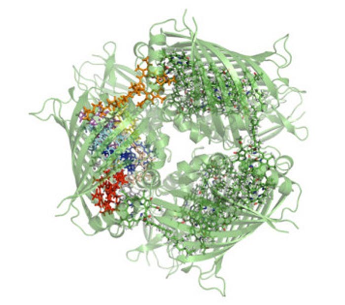
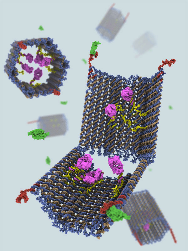
All cells are surrounded by a membrane that controls the flow of substances into and out of the organism. Membranes are made up of molecules called lipids within which proteins are embedded. They were once thought to be essentially uniform, but a number of experiments starting in the 1970s and 1980s indicated that the lipids in fact cluster to form distinct structures tens or hundreds of times larger than the lipid molecules themselves. Scientists did not understand, however, where the energy needed to maintain such structures came from.

In 2008 biophysicist Sarah Veatch at Cornell University in upstate New York and colleagues found a solution. It was known that above 25 °C membranes isolated from live mammal cells exist in a single liquid phase, whereas below that temperature they separate out into two distinct phases, composed of different kinds of lipids and proteins – a bit like oil and water refusing to mix when brought together. What Veatch's group discovered was that as they lowered the temperature of the membranes close to that at which the phases separate out, known as the critical point, small fluctuating patches of the second phase started to appear. Such fluctuations – which measured several microns across and were visible in an optical microscope – do not require large amounts of energy to form.

Critical look at criticality

Veatch has since moved to the University of Michigan but for the current research teamed up with two physicists back at Cornell, Benjamin Machta and James Sethna, to understand the purpose of this criticality. The researchers reckoned that certain kinds of proteins are attracted to one of the phases while other kinds are attracted to the second phase, so tending to draw like proteins together and separate out unlike proteins. As Veatch explains, these interacting proteins would form "signalling cascades" to transmit information regarding the identity of compounds in a cell's vicinity from receptor proteins in the membrane to the inside of the cell. Such information could be used, for example, to decide whether it is a good time to divide or whether it is safe to crawl towards food. "We think that one reason cell membranes contain critical fluctuations is to help facilitate some of the early steps in these signalling pathways," she says.

To calculate the strength and form of the Casimir forces between proteins, Machta used mathematics developed originally for string theory. He found that, as expected, the forces are attractive for like proteins and repulsive for unlike ones, and that they yield a potential energy several times that of the proteins' thermal energy, over distances of tens of nanometres. Much stronger electrostatic interactions, he explains, are limited to ranges of about a nanometre by the screening effects of ions inside the cell. "We have found that by tuning close to criticality, cells have arranged for a long-ranged force to act between proteins," he says.

Sethna adds a broader perspective. "It is amazing how many reactions in cells all involve energies of the same size as thermal fluctuations," he says. "We think that it is the cell being economical – why pay more?"

Something to sneeze at

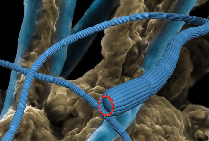
The researchers suspect that the existence of these critical Casimir forces explains why cells low on cholesterol do not function as they should – the removal of the cholesterol, they reckon, taking the membrane away from its critical point. They also speculate that the forces are involved in the sneezing process. Sethna explains that when the receptor proteins in immune cells detect an allergen such as pollen they cluster together, and this clustering somehow triggers the histamines that cause sneezing. He says that perhaps an allergen simply changes the preference of the receptor proteins for one of the two liquid phases in the membrane, hence drawing them together.

The team is hopeful that its work could lead to medical applications. Veatch explains that defects in lipids are thought to contribute to a large number of diseases, including cancer, auto-immunity diseases, and inflammation. "This work may shed light on how lipids could impact some aspects of these diseases," she says. "In the future, I can imagine drugs that specifically target lipids to regulate interactions between proteins in order to treat human disease."

Sethna adds, however, that the time scale for such applications is likely to be long. "Our work is more like figuring out how to make better concrete to build the subbasement of the skyscraper that eventually would house the penthouse of health applications," he says.

Theory explains behaviour

But in addition to any future applications, Sethna argues that the existence of criticality within cells lessens the reliance on purely evolutionary mechanisms when trying to understand how cells operate. "There are lots of things about cells that biologists assume happen because 'evolution made it so'," he says. "Here, I guess, evolution allowed the cell to find this critical point. But once the cell is at the critical point, we can use systematic, cool theory to explain lots of the behaviour, without repeatedly accounting for everything using evolution."

However, some independent experts feel that Veatch's experimental results must be treated with caution because they were not obtained using intact cells. One, who asked to remain anonymous, argues that the separation of the membrane from the rest of the cell might have removed certain relevant components from the membrane and that the body of the cell itself might influence the critical fluctuations in some way. "I am not yet convinced that the theory presented is applicable to in vivo biological membranes," he says. "I therefore think that much more experimental work has to be done to investigate this phenomenon."

The research is described in Physical Review Letters.

Topological quantum computing moves closer

An international team of physicists is the first to implement in the lab an important "error correction" technique that could play a vital role in the development of practical quantum computers. Known as topological error correction (TEC), the technique is based on "clusters" that each contain eight highly entangled photons. These clusters are useful for this purpose because a measurement on one photon does not destroy the entire entangled state.

The multiparticle cluster state at the centre of the current work was first proposed in 2001 by Robert Raussendorf and Hans Briegel, who were then at the University of Munich. Now at the University of British Columbia in Canada, Raussendorf is also involved in this latest research. Such a cluster could be used to perform "one-way" quantum computing, in which the states of individual particles are measured in a specific sequence so that the quantum state of the remaining particles gives the result of the computation.

Like a doughnut

Although quantum computers promise a lot, anyone wishing to build a practical device has to deal with the tricky fact that the quantum nature of qubits fizzles away rapidly as they interact with the heat and noise of the surrounding environment. Quantum error correction offers a way of staving off this "decoherence" – at least long enough for a quantum computation process to occur – by distributing the quantum information held in one "logical" qubit among a number of entangled "physical" qubits. Subjecting these physical qubits to an error-correction algorithm can then reveal if one or more qubits has undergone decoherence and, if so, to restore quantum information.

Developed by Jian-Wei Pan and colleagues at the University of Science and Technology of China in Shanghai, along with Raussendorf and other physicists in Canada and Australia, the new experimental demonstration of TEC involves defining qubits in terms of fundamental shapes that cannot be changed by continuous deformations. A doughnut, for example, remains a doughnut if it is poked, stretched or prodded – unless the perturbation is so violent that it cuts the loop. Topological qubits are similar in the sense that they are not easily perturbed by noise and heat, and must take a big hit before they are destroyed.

The team's cluster state comprises eight entangled photons, each acting as a physical qubit that can have a value of "0" and "1" depending upon its polarization state. The state is made by creating four pairs of entangled photons from firing a laser pulse at a non-linear crystal. The pairs are separated and combined in new pairs that are entangled by having them interfere on polarization-dependent beamsplitters.

The photons can be thought of as forming a 3D cube, in which each photon is entangled with its nearest neighbours. This arrangement has a certain topology that protects a specific quantum correlation between two physical qubits – something that could be used as a building block to create logical qubits in a topological quantum computer.

Repairing qubits

The TEC is implemented on the cluster state by making a series of measurements on the photons – essentially performing a one-way quantum-computing algorithm. To test the correction scheme, the team purposely introduced errors into the system. First, the researchers caused decoherence in one specific qubit and found that the TEC algorithm could identify which photon was affected and correct the error. Next, the team introduced a fixed amount of decoherence to all photons simultaneously, and again the scheme was able to identify the problem and correct it.

"Our experiment provides a proof of principle that topological error correction would be one of the most practical approaches for designing quantum computers," Pan told physicsworld.com.

Pan points out that TEC offers several benefits when compared with conventional schemes – in particular, it can handle the highest error rates of any scheme, making it easier to use with real physical devices, which will always suffer from errors. "Moreover, the architecture used in topological error correction is rather simple: it is sufficient to create interactions between two quantum bits that neighbour each other," he adds. This means that TEC should be compatible with a range of different qubit schemes, including quantum dots and Josephson junctions. This is important because such solid-state qubits should be easier to integrate and scale up to create a practical quantum computer.

Important result

Raymond Laflamme, director of the Institute for Quantum Computing at the University of Waterloo in Canada, says that the work is an important result that shows that TEC can be implemented in principle. But given that not all types of qubits are compatible with TEC, Laflamme cautions that its future usefulness will depend on which qubit technologies are ultimately used to create practical quantum computers.

The next step in the team's research is to create cluster states involving larger numbers of qubits – to do TEC on a logical qubit rather than just a correlation. Ultimately, physicists would like to develop systems that implement TEC on topological qubits and topological quantum-logic gates.


The work is described in Nature

نگراني دانشمندان از كشف تارعنكبوت عجيب در زباله‌هاي هسته‌اي

محققان آمريكايي در حال بررسي تارهاي عنكبوت سفيد رنگ عجيبي هستند كه در استخر زباله‌هاي هسته‌اي كشف شده‌اند. به گزارش پایگاه زیست شناسی ایران، كارمندان بخش زباله هاي هسته‌يي سايت ساوانا ريور در كاروليناي جنوبي ماه گذشته تارهاي عنكبوت عجيبي درعمق 5 و 9 متري استخر سوخت هاي مصرفي پيدا كردند.

چنين ماده سفيد رنگ رشته مانندي تاكنون در بين زباله‌هاي هسته‌اي ديده نشده بود و كشف آن باعث نگراني دانشمندان در خصوص احتمال وقوع جهش ژنتيكي در عنكبوت‌ها شده است. كارشناسان آزمايشگاه ملي ساوانا ريور نمونه‌هاي كشف شده را جمع‌آوري كرده و در حال بررسي دقيق آنها هستند.

اسيديته و دماي آب در استخر سوخت‌هاي مصرفي در اندازه خاصي قرار دارد و در صورت ورود ميكرو ارگانيسم ها به داخل استخر امكان زنده ماندن آنها وجود دارد. حضور هر موجودي در استخر سوخت‌هاي مصرفي، آن را در معرض پرتوهاي راديواكتيو قرار مي‌دهد و احتمال تغييرات ژنتيكي و مبدل شدن به حيوانات بزرگ‌تر را افزايش مي دهد. اگر تارها واقعا توسط عنكبوت‌ها در اعماق استخر درست شده باشند، احتمال قرار گرفتن آنها در معرض پرتوهاي راديواكتيو و جهش ژني عنكبوت وجود دارد.

تنها برخي ارگانيسم‌ها خاصيت مقاومت طبيعي در برابر تابش و پرتوهاي راديواكتيو دارند كه از آن جمله مي‌توان به باكتري «داينوكوكوس راديودورانس» اشاره كرد. اين باكتري يك ارگانيسم طبيعي ضد تابش است كه با استفاده از روش‌هاي مهندسي ژنتيك براي درمان راديواكتيو استفاده مي شود.

DNA nanorobot delivers drugs

Researchers in the US have developed a new nanorobotic device based on DNA that can deliver "cargo", such as drugs, to individual biological cells. The technology might one day be used to treat various diseases by directly programming the immune response of cells.

The nanorobot, developed by Shawn Douglas and colleagues of the Wyss Institute for Biologically Inspired Engineering at Harvard University, US, is in the form of a hexagonal DNA "hinged" barrel that can be opened and closed. The device measures 35 × 35 × 45 nm and can hold various types of cargo – such as metal nanoparticles – in its interior. It is kept closed by two "locks" that are encoded with aptamers – artificial nucleic-acid receptor molecules that bind to specific target molecules, like some antigens – and can be opened like an oyster shell when it interacts with the right combination of antigen "keys". These keys can be proteins on biological cell surfaces and can be made to include specific disease markers, explains Douglas.

Logical locks

The nanorobot can be programmed to open when exposed to a single type of key by using the same aptamer sequence on both lock sites. Another possibility is to encode different aptamer sequences in the locks to recognize two inputs. Both locks need to be opened at the same time to activate the device and it remains firmly shut if only one of the two locks is opened. The lock mechanism thus functions as a logical AND gate: cell-surface antigens either bind ("0") or do not bind ("1") to aptamer locks.

The barrel was made using "DNA origami" in which complex 3D shapes and objects are constructed by folding strands of DNA. Such a technique has already been used to make nano-sized boxes, which are also capable of carrying cargo, with lids that can be locked and unlocked.

"When the nanorobot opens, its previously sequestered biologically active payload can then interact with nearby cells," explains Douglas. "By designing the lock to open when bound to a particular antigen available on a cell surface, the device can thus be targeted to induce cell-signalling 'instructions' in certain cell populations that express the antigen. At the same time, the cargo is prevented from interacting with other cells [that do not express the antigen] in the same environment."

Instructing cancer cells

The researchers have already used their device to deliver instructions, encoded in antibody fragments, to two different types of cancer cells – those responsible for leukaemia and lymphoma. "The instructions were different for both types of cancer cell and contained different antibody combinations," explains Douglas, "and in each case, the message was to activate the cells' 'suicide switch' – a standard feature that allows ageing or abnormal cells to destroy themselves."

The device is the first DNA-origami-based system that employs antibody fragments to convey molecular instructions, something that allows for a completely controlled and programmable way to replicate an immune response, says team leader, George Church. "We are now finally able to integrate sensing and logical computing functions via complex, yet predictable nanostructures," he states. "These are some of the first hybrids of structural DNA, antibodies, aptamers and metal atomic clusters aimed at useful, very specific targeting of human cancers."

The team now plans to test its device on rodents before considering human clinical trials. "Its applications are possibly not just restricted to smart therapeutics but may also be used in diagnostics and even nonmedical applications," adds Church.

شروع پیچیدگی حیات، زیر چشم دانشمندان

براساس مشاهدات پژوهشگران آمریکایی، منشاء حیات چندسلولی که یکی از نقاط عطف تاریخ حیات زمینی‌ به‌شمار می‌رود، می‌توانسته است با سرعتی حیرت‌آور رخ داده باشد. در این پژوهش، یک مخمر تک‌سلولی (موسوم به Saccharomyces cerevisiae)، تنها ظرف مدت ۶۰ روز، به اجتماعات چندسلولی منفرد بدل شد. حتی بعضی از این موجودات چندسلولی، با مرگ خود، راه را برای رشد و تکثیر دیگران فراهم آورده است و این‌گونه یک فرآیند تقسیم کار ساده و ابتدایی را نیز از خودشان نشان دادند.

 
گزارش بررسی مذبور که توسط ویلیام راتکلیف (William Ratcliff) و همکارانش از دانشگاه مینه‌سوتا به ثمر رسیده است، امروز در نسخه آنلاین نشریه Proceedings of National Academy of Sciences انتشار یافت. ریچارد لنسکی (Richard Lenski)، زیست‌شناس فرگشتی دانشگاه ایالتی میشیگان که نقشی در این پژوهش نداشته است، با اشاره به منشأ چندسلولی شدن موجودات می‌گوید: "مدت‌ها چنین چیزی به‌عنوان یک فرایند دشوار تلقی می‌شد، اما این آزمایش‌ها نشان دادند که چندسلولی شدن آنقدرها هم که تصورش می‌رفت، اتفاق سختی نیست."
 
راتکلیف، ایده این آزمایش را به اتفاق همکارش مایکل تراویزانو (Michael Travisano)، طرح‌ریزی کرده بود. او می‌گوید: "داشتیم راجع به جذاب‌ترین کاری که می‌شود انجام داد، فکر می‌کردیم. دیدیم بررسی منشاء حیات کار خیلی سختی است، اما گمان بردیم که درک فرآیند چندسلولی شدن امکان‌پذیر است." این فرآیند، دست کم ۲۵بار به شکل مستقل در طول تاریخ حیات زمین اتفاق افتاده است، اما قدمت همگی‌شان به قدری بالاست که کار بررسی را سخت می‌کند. به‌گفته راتکلیف، "تقریباً غیر ممکن است که با نگریستن به موجودات زنده چندسلولی، پی به ویژگی‌های مربوط به نیاکان بسیار دورشان برد."
 
راتکلیف و تارویزانو، در عوض آمدند و دیدند آیا می‌‌توانند حیات چندسلولی را به کمک چندین موجود تک‌سلولی به‌دست آورند یا نه. آن‌ها نیروی جاذبه را به‌عنوان عامل برگزیننده فرض گرفتند: پیداست توده‌های سلولی، سریع‌تر از نمونه‌های‌ منفردشان در انتهای یک لوله پر از مایع ته‌نشین شدند. راتکلیف، بعد از کشت دادن سلول‌هایی که زودتر ته نشسته بودند، فقط آن‌هایی که چسبیده‌‌به‌هم مانده بودند را برگزید. پس از چندین‌بار گزینش اینچنینی، این سلول‌ها طی بالغ بر ۶۰ روز همچون برف‌دانه‌هایی متشکل از ده‌ها سلول منفرد درآمدند.
 
امنیت در گرو جمعیت
 
اکثر موجودات تک‌سلولی از قبیل مخمرها، غالباً دست به تشکیل اجتماعاتی می‌زنند که در واقع از لحاظ ژنتیکی، همچنان تک‌سلولی به‌شمار می‌روند، اما برف‌دانه‌های راتکلیف، از سلول‌های کاملاً مشابه و به‌هم‌چسبیده شکل یافته بود. بسیاری از دیگر موجودات چندسلولی هم ممکن است از طریق فرآیندی شبیه به همین "تقسیم و الحاق" ایجاد شده باشند.
 
رفتار این برف‌دانه‌ها، درست مثل موجودات چندسلولی واقعی بود. آن‌ها چرخه حیاتی ساده‌ای داشتند که با دوره نوجوانی آغاز می‌شد و بعد از فرآیند رشد بی‌وقفه‌ای که راه به مرحله بزرگسالی‌شان می‌برد، به ابعاد مشخصی می‌رسیدند و به یک بخش بزرگ‌تر موسوم به پدر- مادر و بخشی کوچک‌تر از آن، موسوم به دختر، تقسیم می‌شدند. راتکلیف حتی می‌توانست این چرخه‌ها را به اختیار خودش تنظیم کند: اگر برف‌دانه‌هایی که سریع‌تر ته‌نشین شده بودند را کِشت می‌داد، به چندسلولی‌های بزرگ‌تری هم می‌رسید که پیش از تقسیم‌شان، فربه‌تر می‌شدند. این مسئله، به‌ وضوح نشان داد که انتخاب طبیعی، تأثیرش را به جای اینکه بر سلول‌های منفرد بگذارد، بر مجموعه‌شان اِعمال کرده بود. به‌گفته راتکلیف، "آن‌ها به‌عنوان یک کل می‌زیند و به عنوان یک کل هم از بین می‌روند. در این حالت است که انتخاب، به مرحله چندسلولی منتقل می‌شود".
 
برف‌دانه‌ها تقسیم می‌شوند، چون‌ برخی از سلول‌های سازنده‌شان دست به قربانی کردن خود می‌زنند و بدین‌وسیله به دیگر اجزا، اجازه ترک گروه را می‌دهند. از این‌رو مرگ این سلول‌ها به مصلحت کل جمع است و زمینه رشد برف‌دانه اصلی و تولید فرزندان بیشتر را فراهم می‌کند. این فرآیند، بازتاب‌دهنده تقسیماتی است که در موجودات چندسلولی ِ به مراتب پیچیده‌تر اتفاق می‌افتد، به‌طوری که طی هر نسل، بعضی از سلول‌ها می‌میرند و بدین‌واسطه سلول‌های حاوی اطلاعات ژنتیکی (یعنی اسپرم‌ها و تخمک‌ها)، به فرزندان منتقل می‌شوند.
 
سایر بررسی‌ها در گذشته مشخص کرده‌اند که سلول‌های منفرد، هم از طرفی برای کاهش ریسک شکار شدن‌شان و هم از طرفی برای افزایش شانس دسترسی به غذا، از بودن با یکدیگر سود می‌بر‌ند، اما پژوهش راتکلیف، از معدود نمونه‌هایی است که نشان از نحوه تبدیل اجتماعات تک‌سلولی، به موجودات منفرد و چندسلولی می‌دهد. همایون باقری که در دانشگاه زوریخ سوئیس، به بررسی نحوه تکامل فرآیند چندسلولی شدن مشغول است، می‌گوید: "آن‌ها با مشاهده رخدادهایی که در پی تکمیل فرآیند چندسلولی شدن اتفاق می‌افتد، کار بسیار جالبی را انجام داده‌اند". لنسکی نیز می‌گوید: "گمان کنم این بررسی، نقطه شروعی برای کارهای واقعاً مهمی است که سال‌ها طول می‌کشد تا به ثمر بنشینند؛ کارهایی که نه فقط نشان از زیرلایه‌های ژنتیکی این ماجرا می‌دهند، بلکه مدت‌زمان دوام مخمرها در حالت چندسلولی‌شان را هم تعیین می‌کنند.
 
مخمرها، خود از اجداد چندسلولی پدید آمده‌اند و لذا بعید نیست به‌همین‌واسطه در مدت‌زمانی چنین کوتاه موفق به بازسازی سبک زندگی باستانی‌شان ‌شده‌ باشند. با این همه، راتکلیف اعتقاد دارد که مخمرها، چندین‌میلیارد نسل پیش، از موجوداتی چندسلولی به تک‌سلولی بدل شدند و در همان اوقات هم ژن‌های مربوط به چندسلولی‌شدن را از دست داده‌اند، اما او می‌خواهد همین آزمایش را این‌بار روی تک‌سلولی‌هایی نظیر جلبک‌های سبز دریایی (از تیره Chlamydomonas ها)، که پیشینه چندسلولی نداشته‌اند، انجام دهد.
 
به نقل از Nature

راه رفتن ما، میراث ماهیان ریه‌دار

ماهی‌هایی هستند که جست‌وخیزکنان، در حالی‌که بر باله‌های خود ایستاده‌اند، از آب بیرون می‌زنند و حین پیاده‌روی‌شان روی خشکی، از هوای آزاد آن تنفس می‌کنند. به‌گفته دانشمندان، از روی رفتار همین ماهی‌ها شاید بتوان گفت که راه رفتن، قبل از مهاجرت آبزیان به خشکی، در زیر آب در عمل به تکامل رسیده است. می‌توان گفت اجداد کهن انسان‌ها و تمامی پستانداران، خزندگان، پرندگان، دوزیستان و سایر چهارپایان، در واقع ماهیانی هستند که در نهایت توانایی تنفس روی خشکی را به دست آورده‌اند. یکی از اندک‌گونه‌های بقایافته از این تیره‌های کهن خشکی‌زی، جانوران هوازی و کمیابی موسوم به "ماهی ریه‌دار" (Lungfish) هستند که امروزه در مناطقی از آفریقا، آمریکای جنوبی و استرالیا زندگی می‌کنند.

حال، دانشمندان متوجه شده‌اند که ماهی ریه‌دار آفریقایی (با نام علمی Protopterus annectens) می‌تواند به کمک باله‌های لاغرش، بدن خود را به تمامی از روی زمین بلند کند و به پیش بیندازد. این قابلیتی است که تا پیش از این، تصور می‌رفت از رفتارهای چهارپایان نخستین – که مدت‌ها پس از این ماهیان ظاهر شده بودند – پدید آمده باشد.
 
نیل شوبین (Neil Shubin)، پژوهشگری از دانشگاه شیکاگوست که می‌گوید: "رفتار این ماهی‌ها، نشانگر گام‌هایی‌ست که منشاء راه رفتن بوده‌اند. آنچه ما در ماهی ریه‌دار می‌بینیم، نمونه‌ای فوق‌العاده زیباست که به ما نشان داده چگونه راه رفتن روی دوپا در ماهیان آب‌زی، آسان و بی‌دردسر به الگوی راه رفتن در چارپایان منتهی شده است." ماهی ریه‌دار مد نظرما، بدنی شبیه به مارماهی مجهز به یک جفت باله سست و بی‌دوام دارد. "اگر اسکلت این جاندار را به من نشان داده و با من شرط ببندید که راه می‌رفته یا نه، شرط می‌بستم که نمی‌توانسته راه برود." این را شوبین می‌گوید و می‌افزاید: "باله‌هایشان به همه چیز شباهت دارد، الا اندامی برای راه رفتن."
 
تاریخچه غنی حیات ماهیان ریه‌دار، این جانوران را به سوگلی‌های دیرینه‌شناسان بدل کرده و سال‌ها بوده که حکایات و شایعاتی هم مبنی بر اینکه رفتاری شبیه به راه رفتن را می‌شود در این ماهیان مشاهده کرد، در بین دانشمندان دهان‌به‌دهان می‌چرخیده است. پژوهشگران، با هدف کشف حقیقت احتمالی پنهان در پس این شایعات، دست به طراحی آکواریوم ویژه‌ای زدند که با کمکش قادر به تصویربرداری از حرکات این ماهی، از زاویه پایین و همچنین از طرفین بودند. فیلم‌های به‌دست‌آمده نشان از این می‌داد که ماهی ریه‌دار، اغلب از باله‌های پشتی‌اش برای خیز رفتن بهره می‌گیرد و همزمان از هر دو باله‌اش برای جهیدن استفاده می‌کند و برای راه رفتن هم حرکات باله‌ای‌اش را به تناوب تغییر می‌دهد.
 
هدر کینگ (Heather King)، زیست‌شناس فرگشتی دانشگاه شیکاگو می‌گوید که "چقدر جذاب و شگفت‌انگیز است که حتی با چنین باله‌های لاغری هم این ماهی ریه‌دار، نه‌تنها می‌تواند خود را به پیش براند، بلکه قادر به بلند کردن کامل بدنش [از کف ظرف] هم هست. اگر قرار باشد که صرفاً به اسکلت این ماهی بنگریم، هرگز حدس هم نخواهید زد که امکان نمایش چنین رفتاری را داشته باشد، علی‌الخصوص از آنجایی‌که این ماهی اصلاً پا ندارد." به‌رغم اینکه باله‌های جلویی ماهی دقیقاً شبیه به باله‌های عقبی‌اش است، اما برای جست و خیز و راه رفتن از آن‌ها بهره‌ای نمی‌گیرد. البته پژوهشگران شک دارند که واقعاً این‌گونه باشد، اما به‌گفته کینگ، "امکان دارد اگر باله‌های عقبی، تنها برای به پیش راندن ماهی کفایت کنند، نیازی به باله‌های جلویی احساس نشود".
 
به‌گفته پژوهشگران، توانایی ماهی ریه‌دار برای حمایت از وزن بدنش روی چنین اندام ظریف و نازکی، احتمالاً به کمک شناور کردن خود از طریق انباشت ریه‌ها با هوا امکان‌پذیر می‌شود. شوبین می‌گوید: "این نمونه به ما نشان می‌دهد در محیط آب که دیگر نبایستی تمامی وزن بدن را تحمل کرد، چه گزینه‌هایی برای سهولت در حرکت پیش روی‌تان است." ممکن است این کشف، مسیر فرگشتی ویژه‌ای را که دانشمندان گمان می‌کردند حیات از طریق آن از آب به خشکی راه پیدا کرده است را دچار دگرگونی‌هایی کند. کینگ مدعی است اغلب مراحل لازم برای وفق دادن خود با محیط خشکی، می‌توانسته میلیون‌ها سال پیش از اینکه چارپایان صاحب پاهای خود شوند و عزم خشکی کنند، رخ داده باشد. این یافته‌ها ممکن است حتی ما را به تجدید نظر در خصوص ماهیت رد پاهای تقریباً ۳۸۰ میلیون‌ساله‌ای رهنمون شود که اخیراً پیدا شده است. رد پاهایی که می‌توانسته اصلاً توسط گونه‌هایی از ماهیان ایجاد شده باشد. کینگ، شوبین و همکاران‌شان، یافته‌های خود را دیروز در نسخه آنلاین نشریه علمی Proceedings of National Academy of Science انتشار دادند.
 
ترجمه از "احسان سنایی"

انرژی مغز از کجا می‌آید؟

قریب به نیم‌میلیون سال پیش، مغز ما انسان‌ها شروع به رشد کرد و مغز بزرگ‌تر هم طبعاً به انرژی بیشتری برای دوام آوردن نیاز دارد، اما دانشمندان از همین متعجب هستند که وقتی‌ آهنگ سوخت و ساز بدن انسان که تأمین‌گر انرژی‌اش است، با عموزاده‌های‌مان که مغزی به ابعاد یک نخود داشتند هیچ تفاوتی نمی‌کند، پس این مقادیر اضافی انرژی از کجا آمده است؟

بر اساس یکی از فرضیات اخیر، نیاز انرژی مغزمان توسط روده کوچک تأمین می‌شود، چراکه غذایی که راحت‌تر هضم می‌شود، انرژی روده را برای رشد مغز آزاد می‌کند، اما پژوهش‌های تازه مدعی‌اند که با توجه به اهمیت بیشتر انرژی موجود در ذخیره چربی بدن، این فرضیه ممکن است اصلاً صحیح نباشد. "حیواناتی که مغز بزرگ‌تری دارند، از بافت‌های نازک‌تر چربی هم برخوردارند و حیواناتی که بافت چربی‌شان برتری دارد، ابعاد مغزشان کوچک‌تر است." این را آنا ناوارته (Ana Navarrete) از دانشگاه زوریخ سوئیس می‌گوید و می‌افزاید: "یا شما مغز بزرگ‌تری دارید و یا بافت غنی‌تری از چربی. معمولاً این‌ دو در نسبت معکوس با همدیگرند."
 
مغز، در حدود ۲۲ برابر یک ماهیچه انرژی نیاز دارد تا هر دو با ظرفیت به نسبت برابری واکنش نشان دهند و این انرژی هم از غذایی که می‌خوریم تأمین می‌شود. ابعاد مغز ما انسان‌ها در حدود سه برابر ابعاد مغز نزدیک‌ترین نمونه‌های جانوری به ما، یا شامپانزه‌ها هست و مصرف انرژی‌اش سه برابر بیشتر است، حال آن که نرخ سوخت و ساز هر دو گونه مشابهت دارد. پس این انرژی اضافی، باید از جایی تأمین شده باشد و ناوارته و همکارانش هم برای یافتن این‌جا، دست به بررسی ۱۹۱ نمونه از یکصد پستاندار مختلف زدند. هدف این بود که فرضیه "بافت غنی" (که می‌گفت این انرژی از پس‌مانده‌های ذخیره انرژی روده کوچک به‌دست می‌آید) را به مصاف فرضیه‌‌ای دیگر بفرستند که انرژی مزبور را به غنای بافت چربی بدن نسبت می‌داد. نتیجه آن شد که نسبت مستقیمی نه مابین ابعاد مغز و روده، بلکه بین مغز و میزان چربی موجود در بافت‌های بدن جانداران پیدا شد و در جانواران وحشی و جنس ماده نیز از همه بارزتر بود. به‌علاوه، این نسبت برای ۲۳نمونه اول اصلاً صدق نمی‌کرد، شاید از آنجا که همگی این نمونه‌ها، مربوط به حیوانات به اسارت درآمده‌ای بودند که ذخیره چربی‌شان یا بیشتر یا کمتر از هم‌خانواده‌های وحشی‌شان بود.
 
ذخیره بالای چربی، باعث کاهش سرعت جاندار حین حرکاتی از قبیل صعود به ارتفاع، پرواز یا فرار از دست صیادان می‌شود، اما ذخیره انرژی خوبی هم به‌شمار می‌رود. این مسئله حکایت از این می‌کند که به‌گفته ناوارته، بقای جاندار در گرو دو راهبرد است: یا برای اوقات دشوار زندگی، چربی بیشتری که به‌معنای ذخیره انرژی بیشتری هم می‌شود ذخیره کن، یا مغز بزرگ‌تری داشته باش و به فکر رهایی از این ناملایمات بربیا.
 
اما انسان‌ها، هم مغز بزرگی دارند و هم چربی فراوان و این بدین‌معناست که در ناملایمات زندگی، برای بقا به هر دو گزینه می‌توان متکی بود. ناوارته مدعی‌ست که چیز دیگری هم در این میان از قلم افتاده که به‌نحوی مربوط به رهیافت بهینه ما انسان‌ها در امر تحرک می‌شود. هرچند که او نمونه‌‌های آزمایشگاهی مربوط به انسان را مورد بررسی قرار نداده، ولی معتقد است که ما به‌واسطه سبک کاملاً متفاوت جابجایی‌مان نسبت به سایر جانداران، عادات پیشین حرکت را شکسته‌ایم. راه رفتن روی دو پا، نسبت به پرسه زدن با هر چهار دست و پا در لابه‌لای درختان همانند شامپانزه‌ها، از حیث مصرف انرژی، فرآیند جابجایی به‌صرفه‌تری‌ محسوب می‌شود. پس از آنجایی که داشتن ذخیره چربی برای ما انسان‌ها گران تمام نمی‌شود، هم می‌توانیم از این ذخیره بهره بجوییم و هم اینکه از مغزمان برای یافتن منابع غذایی کمیاب‌تر بهره بگیریم.
 
با این‌ وجود، نبود نمونه‌های انسانی در این پژوهش، برخی از دانشمندانی که در آن نقشی نداشته‌اند را نگران کرده و می‌گویند امکان دارد تفاسیر ناوارته، مبالغه‌آمیز باشد. جک بیکر (Jack Baker)، پژوهشگری از دانشگاه نیومکزیکو که در این پژوهش هیچ نقشی نداشته است، می‌گوید: "این نتایج حاکی از آن است که در انسان‌های نخستین، ذخیره چربی، به نسبت رشد ابعاد مغز اصلاً فدا نشده است. اهمیت این پژوهش، صرفاً حول محور ارتباط مابین این نتایج و فرضیه بافت غنی می‌چرخد – که تقریباً مختص خاستگاه انسان است. حال‌آنکه در این پژوهش هیچ نمونه‌ای از انسان مورد بررسی قرار نگرفته است." لزلی آیلو (Leslie Aiello)، پژوهشگری از بنیاد Wenner-Gren نیویورک هم که در این پژوهش نقشی نداشته است، می‌گوید: "با این‌حال، ناوارته و همکارانش، مجموعه داده‌های بی‌سابقه‌ای را گرد آورده‌اند که پیشرفت چشمگیری نسبت به آنچه ۲۰ سال پیش در اختیارمان بود، به حساب می‌آید."
 
به‌گفته آلیو، این مجموعه داده‌ها، اطلاعات بیشتری را به معمای تکامل مغز آدمی می‌افزاید، اما سئوالات و پیچیدگی‌هایی را هم ایجاد می‌کند که پاسخ واحدی نمی‌شود به آن‌ها داد و باید از مجموع دانسته‌های‌مان درباره ابعاد روده کوچک، نسبت بالای ذخیره چربی انسان، سبک خاص جابه‌جایی‌مان و دیگر مؤلفه‌های قابل بحث، پاسخی بیرون کشید. این پژوهش، در نشریه علمی nature منتشر شده است.

Calcium ions simulate the quantum world

The first digital "quantum simulator" based on trapped ions has been built by physicists in Austria. The system, developed by Ben Lanyon and colleagues at the University of Innsbruck, comprises a number of trapped calcium ions that are manipulated using sequences of laser pulses. The team has used the system to simulate the time-evolution of several multi-particle systems.

A quantum simulator uses one quantum system to simulate the behaviour of another, less accessible system. For example, by carefully manipulating the laser light and magnetic fields trapping an ensemble of ultracold atoms, researchers can control the interactions between atoms – and therefore simulate interactions that occur between electrons in solids. But unlike electrons in solids, the strength of these interactions can be easily adjusted, allowing physicists to test theories of condensed-matter physics.


Analogue to digital

Most quantum simulators are "analogue" in the sense that the interactions between the trapped atoms are directly analogous to those between electrons. A digital quantum simulator, in contrast, contains an ensemble of interacting quantum particles that act as quantum bits (qubits) and can be used to create quantum logic gates. The quantum system to be simulated is then encoded into the system and the behaviour of the electrons is determined by performing a quantum calculation.

Unlike analogue simulators, which address specific systems, a digital simulator could be used to study a wide range of quantum systems. Furthermore, digital simulators can benefit from error-correction schemes, which means that physicists can be more confident in their results.

But while researchers have had some success creating digital quantum simulators using nuclear magnetic resonance (NMR) techniques, these work with just two or three qubits and it has proven difficult to scale up to the 40 or so qubits needed to do a useful quantum simulation. The new trapped-ion quantum simulator created Lanyon and colleagues means that it should, in principle, be much easier to scale up such a system to do useful simulations.

Easily scalable

The team's experiments begin with a small number of calcium ions (a maximum of six) that are lined up in a row in an electromagnetic trap. Each ion can exist in two electronic states – "0" and "1" – and can therefore act as a qubit. Interactions between individual ions can be controlled by firing carefully selected laser pulses at the trapped ions.

A calculation begins by putting the ions into a specific quantum state. In an experiment involving four ions, for example, each qubit was given the value "1". A series of laser pulses was then fired at the ions, which causes them to interact with each other creating a sequence of logic gates that process the quantum information held in the initial state.

It is this sequence that simulates the interactions that occur in a real (or imagined) quantum system. In this particular example, the qubits were used to simulate four spin-1/2 particles in which the spin of each particle can interact with the three other particles.

Approximate solutions

Lanyon and colleagues were interested in calculating the time evolution of the spins, which is particularly difficult to do using a classical computer. To do this, the team implement the "Trotter approximation" on their system. This is done by firing a series of pulses that simulates the evolution of the system over a certain period of time before the values of the qubits are read out. The system is then reset and an identical simulation is repeated many times to obtain average values for the qubits – which is an approximate solution to the problem being simulated.

This entire process is then repeated to simulate a number of different time periods, building up a map of the time evolution of the spins. Time evolution simulations were done for as many as six trapped-ion qubits and involving up to 100 quantum gates.

Towards quantum chemistry

"Six qubits and a 100 gates for quantum simulation is a feat that paves the way for more complex and rich digital quantum simulations in the future," says Alán Aspuru-Guzik of Harvard University in the US. "What Ben Lanyon and his colleagues did was to implement one of the most important building blocks for quantum simulation, what we call a 'Trotter step' in a generic or universal sense. This is one of the required and essential building blocks to do exact quantum chemistry on quantum computers, when they become powerful enough."

Lanyon told physicsworld.com that his team's next challenge is to perform the simulations with 10 or more ions. Creating such a system is not the problem – the team has already trapped and entangled as many as 14 ions. However, performing large numbers of operations on the ions is tricky because the qubits tend to lose their quantum nature over time as they interact with their surroundings.

Physicists in tune with neurons

Have you ever wondered why certain sets of musical notes sound perfectly melodious while others make you want to cover your ears? Now, physicists in Europe have developed a model that suggests that certain notes sound harmonious because of the consistent rhythmic firing of neurons in the auditory system. The researchers say that they have now quantified this effect by showing that the neural signals are regularly spaced for frequencies that are pleasant sounding, but are erratic for those that are not. They say that their model may also provide insights into other senses, such as vision, that employ similar neural processing systems.

Hitting the right notes

How humans and animals perceive sound has long fascinated scientists because of the brain's amazing ability to process auditory signals. "The auditory sensory system is the most investigated apparatus. But even simple sound signals – pairs of pure tones such as musical chords – are able to cause phenomena that can not be simply explained," says Bernardo Spagnolo of the University of Palermo, Italy, and one of the authors of the study published in Physical Review Letters this month.

As examples, Spagnolo cites two things that musicians take for granted: the "pitch perception" and the "perception of consonance and dissonance". Simply put, perceiving pitch is simply being able to differentiate clear frequencies of sounds, while the perception of consonance and dissonance is being able to tell the difference between harmonious and inharmonious chords. In their current research, the researchers focused on consonance and dissonance perception with the aim of identifying the location and quantity of the signals associated with harmony and disharmony in the brain.

On board the "spike trains"

Hearing involves the conversion of sounds into neural "spike trains". The researchers' model comprises three neural-like elements. Two of these elements represent sensory neurons and are driven by noise at two different frequencies. The outputs of two neurons, in the presence of a "noisy" environment, are synaptically connected with the third neuron, which represents an "interneuron". This is an internal neuron that connects a sensory neuron to other neurons in the brain. In reality there are more than two initial neurons, as the human ear can operate at frequencies of between 20 Hz and 20 kHz and can detect sounds over a range of 120 decibels.

The output spike train of the interneuron is the main focus of the study, which found that if an acoustic signal is transformed by the auditory system into spike trains with a regular distribution of inter-spike intervals, then the signal is perceived as harmonious. But when the inter-spike intervals are irregular, the signal is perceived as inharmonious. At the output of the interneuron, inharmonious input signals give rise to blurry spike trains, while the harmonious signals produce more regular, less "noisy" spike trains.

Senses and sensibility

The research team quantified the regularity of the interneuron output in terms of the entropy of the signal. "This regularity is linearly connected with informational entropy: harmonious chords give rise to high spike-train regularity and so low entropy; inharmonious chords give rise to low spike-train regularity and so high entropy."

Spagnolo points out that the model can investigate the role of external and internal "environmental noise" in the nervous system, with respect to the sensory phenomena of "recognition" and "permanence of information" contained in complex input signals in the brain. "Investigating this process can help to understand which types of input signals are able to survive in the noisy environment of the brain, reveal the mechanism of this process, and explain what it means from a perceptional and cognitive point of view," claims Spagnolo. He also says that studying and understanding the auditory system provides a basis for other less-studied sensory systems "that exhibit the analogous principles of conversion of environment stimuli into the neural spike trains".

نانوسیم های زیگزاگی حرکت براونی را محدود می کنند

فیزیکدانان در ایالات‌متحده تله مغناطیسی ساخته‌اند که می‌تواند ذرات میکروسکوپی را علی‌رغم حرکت براونی آن‌ها به دام اندازد. این تله که بر مبنای نانوسیم‌های مغناطیده با شکل زیگزاگی قرار دارد، می‌‌تواند به محققان در انجام آزمایش‌های شیمیایی و زیست‌شناسی در محیط ریزسیال کمک کند؛ در چنین محیطی سیالات به لحاظ هندسی مقید به مقیاس زیرمیلیمتری هستند.

ریزسیالش(Microfluidics)، حوزه‌ای نوظهور است که شامل انتقال پیکولیتری مایعات از طریق تونل‌هایی با پهنای میکرونی است. توانایی اجرای ازمایش‌ها در واحدهای کوچک، برای بسیاری از پژوهشگران شیمی، زیست‌شناسی و پزشکی مفید است زیرا مجبورند با موادی کار کنند که ترکیب آن‌ها - همانند داروهای جدید - گران یا دشوار است. به‌علاوه، چندین سامانه ریزسیال جدید می تواند با هم تعامل کرده و «آزمایشگاهی روی تراشه» بیافریند که پایه ای برای مطالعات بسیاری از فرایندهای شیمیایی باشند.

نیاز کلیدی ریزسیالش و نانوفناوری به طور عام، توانایی انتقال اجسام در مسیرهایی با طول 100 نانومتر تا 10 میکرومتر است،جایی که افت و خیز های تصادفی و حرکات گرمایی که «حرکت‌ براونی» نامیده می شوند، نقش بزرگی بازی می‌کنند. بدین منظور فنون متفاوتی ابداع گردیده اما هر کدام نواقصی دارند. برای مثال انبرک‌های نوری می‌توانند ذرات را با میدان‌الکتریکی تولید‌شده توسط یک پرتو لیزر متمرکز به دام بیندازند اما این فرایند می‌تواند باعث گرمای موضعی شود. انبرک ‌های دی‌الکتریک با میدانی الکتریکی بین الکترودها، نیز محیط را به شدت مختل می‌کنند.

فرمان های زیگزاگی مغناطیده

اکنون، هارون چن(Aaron Chen) و همکارانش در دانشگاه ایالتی اهایو در کلمبوس ایالت متحده، تله‌ی ذراتی را معرفی نموده‌اند که ممکن است راهی برای برون‌رفت از این مشکلات داشته باشد. تله شامل یک سیم مغناطیده، ساخت‌ شده از آهن و کبالت است. پژوهشگران روی یک سطح سیلسیومی آن را به شکل زیگزاک در آورده اند. آنها ابتدا یک میدان مغناطیسی قوی اعمال کردند و در‌نتیجه نقاط خمیده در راس ها به شدت مغناطیده شده و هر راس شبیه یک تک قطبی مغناطیسی با میدانی که به آن وارد میشود یا از آن خارج میشود رفتار میکند. راس‌ها به این گونه مانند تله های مغناطیسی عمل می کنند. سپس با اعمال میدان‌مغناطیسی ضعیف‌تر، قدرت تله را میزان کرده و این گونه رو رفتار ذرات را کنترل می‌کنند.

در این آزمایش ذرات دی‌اکسید آهن کپسول شده در یک پلیمر مورد استفاده قرار گرفته اند که شعاع کل آنها از 0.28 تا 0.6 میکرومتر است. این ترکیب به ذرات خاصیت ابرپارامغناطیسی می بخشد و در نتیجه می توانند در میدان‌های مغناطیسی نسبتا ضعیف نیز مغناطیده شوند بدون این که مغناطش دایم پیدا کنند. با استفاده از دوربین CCD، محققان دیدند که ذرات در تله ای در حدود 100 نانومتر قرار دارند. به عبارت دیگر، تله می تواند بر حرکت براونی ذرات بدون اتصال کامل به آنها فایق آید. چن و همکارانش اکنون می‌خواهند روش خود را برای تله انداختن بیش از یک ذره گسترش دهند. او می گوید:«بررسی چگونگی [حرکت] و برهمکنش ذرات درون تله، هدف اصلی بعدی ماست.»

لینک منبع مقاله

Radioactive decay accounts for half of Earth's heat

About 50% of the heat given off by the Earth is generated by the radioactive decay of elements such as uranium and thorium, and their decay products. That is the conclusion of an international team of physicists that has used the KamLAND detector in Japan to measure the flux of antineutrinos emanating from deep within the Earth. The result, which agrees with previous calculations of the radioactive heating, should help physicists to improve models of how heat is generated in the Earth. Geophysicists believe that heat flows from Earth's interior into space at a rate of about 44 × 1012 W (TW). What is not clear, however, is how much of this heat is primordial – left over from the formation of the Earth – and how much is generated by radioactive decay.

The most popular model of radioactive heating is based on the bulk silicate Earth (BSE) model, which assumes that radioactive materials, such as uranium and thorium, are found in the Earth's lithosphere and mantle – but not in its iron core. The BSE also says that the abundance of radioactive material can be estimated by studying igneous rocks formed on Earth, as well as the composition of meteorites.

As a result of this model, scientists believe that about 20 TW is generated by radioactive decay – 8 TW from the uranium-238 decay chain; 8 TW from the thorium-232 decay chain and the final 4 TW from potassium-40. Fortunately, these decay chains also produce anti-electron-neutrinos, which travel easily through the Earth and can be detected, thereby giving physicists a way to measure the decay rates and ultimately the heat produced deep underground.

Decay and measure

In 2005 researchers at KamLAND announced that they had detected about 22 such "geoneutrinos", while last year scientists at the Borexino experiment in Italy said they had detected 10. Now, the KamLAND team has bagged a total of 111 of these tiny almost massless particles. The combined results have allowed the KamLAND team to conclude that the heat flux due to the uranium and thorium decay chains is about 20 TW with an uncertainty of about 8 TW. While the KamLAND experiment cannot detect the lower-energy antineutrinos from potassium-40 decay, the researchers believe that the value predicted by the BSE model of 4 TW is correct.

Although 20 TW from uranium and thorium is more than the 16 TW predicted by the BSE model, it is still within the experimental uncertainty – and is much less than the total flux of 44 TW. "One thing we can say with near certainty is that radioactive decay alone is not enough to account for Earth's heat energy," says KamLAND collaborator Stuart Freedman of the Lawrence Berkeley Laboratory in California. "Whether the rest is primordial heat or comes from another source is an unanswered question."

One possibility that has been mooted in the past is that a natural nuclear reactor exists deep within the Earth and produces heat via a fission chain reaction. Data from KamLAND and Borexino do not rule out the possibility of such an underground reactor but place upper limits on how much heat could be produced by the reactor deep, if it exists. KamLAND sets this limit at about 5 TW, while Borexino puts it at about 3 TW.

Oil-filled balloon

The KamLAND detector is a huge balloon filled with 1000 tonnes of mineral oil that is monitored by more than 1800 photomultiplier tubes. It is located deep underground in a Japanese mine to shield the detector from cosmic rays. Very occasionally an antineutrino will react with a proton in the oil to create a neutron and a positron. The positron travels a short distance through the oil, giving off a flash of light as it ionizes oil molecules. The positron then annihilates with an electron to create two gamma-ray photons. These two processes happen very quickly and the light can be detected by the photomultiplier tubes. In addition, the energy of the antineutrino can be estimated from the amount of light given off during ionization.

A few hundred milliseconds later, the neutron is captured by a proton to form a deuteron. This results in the emission of a gamma ray, which can also be detected by the photomultiplier tubes. By looking for signals in the photomultiplier tubes that are separated by the appropriate amount of time, KamLAND can discriminate between extremely rare antineutrino events and the much more common signals due to background radiation.

اوریگامیِ DNA، انقلابی در تولید فرا ماده

اولین فرا‌ماده برای منحرف کردن امواج میکرویو به شیوه‌های که مواد معمولی توانایی انجام آن را ندارند، در سال 2003 طراحی شد. این ماده از قطعات C شکل فلزی و سیم‌هایی که در ساختار لانه زنبور بهم متصل می‌شوند، ساخته ‌شده است. اندازه این فرا‌ماده مهم است. اِلمان های فعال در فرا‌ماده و ساختار تکراری آن باید کوچک‌تر از طول موج نوری باشد که برای نفوذ به آن طراحی شده‌است. به همین دلیل قطعات C شکل فلزی که فیزیک‌دانان آنها را تشدید‌گرهای حلقوی دو تیکه می‌نامند در کل چند میلیمتر هستند. یعنی برای اینکه بتوان ساختار کلی آنرا بدون زحمت و با دست سرهم بندی کرد، به حد کافی بزرگ هستند.

این مساله پرسش روشنی را پیش می‌آورد: چگونه می‌توان قطعاتی مشابه ساخت که در طول موج‌های کوچک‎‌تر کار کند. برای نور فروسرخ، اِلِمان‌های فعال باید بشکل ساختار تکرار شونده در مقیاس چند صد نانومتر طراحی شوند که با روش‌های لیتوگرافی امروزی کار دشواری نیست.

ولی برای نور مرئی، ساختار تکرار شونده باید از مقیاس چند نانومتر باشد. ساخت چنین چیزی سخت‌تر است. گروه‌هایی هستند که چنین ماده‌ای را با استفاده از نانو میله های بافته شده‌ی تخت (دو بعدی)، ساخته‌اند. اما گام بعدی که ساخت قطعات سه بعدی است بسیار دشوار‌تر می‌شود. این به علت دشواری ساختن اِلِمان‌ها نیست، ساخت نانو ذرات فعال نوری که چند نانومتری هستند راحت است. بلکه سوار کردن آنها بشکل ساختاری سه بعدی، مانند لانه زنبور در مورد فرا مواد میکرویوی، قسمت دشوار کار است.

حالا آنتون کوزیک و دوستانش در دانشگاه صنعتی مونیخ با استفاده از روشی به نام اوریگامیِ دی اِن اِی راهی برای حل این مساله یافته‌اند. ایده آنها این است که نانو ذرات طلا را بوسیله تک رشته‌های کوچک دی اِن اِی پوشش دهند. از طرف دیگر تک رشته‌ی مکمل این رشته ها در ساختار بزرگتری از دی اِن اِی اصطلاحا داربست نامیده می‌شود، قرارداده میشود. هنگامیکه نانوذرات و داربستِ دی اِن اِی در کنار همدیگر در حلال قرارداده می‌شوند، رشته‌های دی اِن اِی مکمل به یکدیگر میچسبند و با این روش نانو ذرات را به داربست می‌چسبانند. اگر طراحی با دقت انجام شود، فن‌آوری اوریگامی دی اِن اِی توانایی ساختن هر شکلی را دارد.

کوزیک و همکارانش از این فرآیند برای پیوند دادن 9 نانو ذره طلا تنها به طول10 نانومتر در طول رشته‌های دی اِن ِای برای ساختن شکل مارپیچی استفاده کرده‌اند. بنابراین ذرات بصورت پله های مارپیچی شکل می‌گیرند و از آنجا که این فرآیند خود‌سازمانده است، این شکل دهی بصورت موازی ادامه می‌یابد‌. در نتیجه توانای ساخت میلیون‌ها پله مارپیچی را بطور همزمان دارد. این فرآیند بطور شگفت آوری تا 80 درصد دقیق است و یک مارپیچ کامل را می‌سازد.

نتیجه یک شاره است که ویژگی‌های نوری ساختار‌های نانو ذره مارپیچی را دارا است. هر نور قطبیده دایروی که درون مارپیچ حرکت می‌کند می‌تواند امواج الکترونیکی که پلاسمون نامیده می‌شود را روی سطح نانو ذرات طلا برانگیخته کند. اگر جهت چرخش نور قطبیده دایروی با جهت مارپیچ هماهنگ باشد، در برخورد با نانوذرات پشت سرهم جذب می‌شود. اما نور با جهت چرخش مخالف مستقیما از آن رد خواهد شد. بنابراین شاره نور با جهتگیری خاصی را جذب می‌کند. به این پدیده دورنگی دورانی می‌گویند.

این دقیقا همان چیزی است که کوزیک و همکارانش مشاهده کردند و معتقدند که می‌توانند این اثرات را به دو صورت تنظیم کنند؛ یک، بوسیله تغییر جهتگیری داربست دی اِن اِی و دو، با رشد دادن لایه نقره روی نانو ذرات طلا که بسامد نوری که به آن حساس هستند را نیز تغییر می‌دهد. آنچه این گروه انجام داده‌اند پیشرفتی مهمی بشمار می‌رود زیرا نخستین بار است که کسی موفق به طراحی فرا ماده یا فرا شاره فعالِ نوری در مقیاسِ بزرگ شده‌است.

این گروه امیدوار به اختراعات بلند پروازانه‌تری هستند و معتقدند با استفاده از روشی که پیشنهاد داده‌اند می‌توان به ساخت موادی با ضریب انکساری منفی که کاربرد‌هایی مانند نامرئی کردن و ساخت لنز‌های بی‌عیب را ممکن می‌کند، امیدوار بود. آنها بر این باورند که روش اوریگامی دی اِن اِی امکان طراحی ساختارهای بسیار پیچیده تر را فراهم می‌کند. این ساختار ها حتی قابلیت تبدیل شدن به جامد را به کمک روش های تبلور دارند.  همه اینها اما ساده نخواهد بود. اوریگامی دی اِن اِی روشی نو و در حال پیشرفت است و تولید بلورِ با کیفیت بالا هرگز آسان نبوده ‌است. اما این باعث توقف تلاش‌های آنها نخواهد شد. زودیک و دوستانش بر این باورند که کارهایشان راه را برای تولید نوع تازه ای از فرا موادِ نوری هموار خواهد کرد. باید منتظر نمونه های بیشتری به‌همین زودی باشیم.

لینک منبع مقاله

ساخت پوستی از جنس مدارهای الکترونیکی

پوست الکترونیکی، فن آوریی برای ثبت ضربان قلب، فعالیت های الکتریکی مغز و انقباض های ماهیچه ای با دقتی مشابه الکترود های حجیم معمولی است. این قطعه که ضخیم تر از تار موی انسان نیست، بوسیله ی یک متخصص علوم مواد در دانشگاه ایلینویز به نام جان راجرز و همکارانش ساخته شده است و شامل شبکه قابل انعطاف و کشسانی از مدار های حسگر است. آن را می توان مانند خالکوبی موقت روی پوست قرار داد و یا از پوست جدا کرد و برای چسباندن به پوست نیازی به چسب ندارد. در حال حاضر از این پوسته ها می توان برای چند روز استفاده کرد، اما پژوهش گران امیدوارند این فن آوری روزی به پزشکان اجازه دهد تا وضعیت سلامت بیماران را بطور دائم زیر نظر بگیرند.


پوست الکترونیکی توانایی انجام کارهایی را دارد که حسگرهای پزشکی مرسوم قادر به انجامشان نیستند. برای مثال هنگامیکه این قطعه در دهان قرار داده می شود کلماتی که بیان می شود را در حدی مناسب برای کنترل یک بازی کامپیوتری ساده، دریافت می کند. به گفتهء راجرز دلیل امتحان آنها روی دهان اینست که نشان دهند که این قطعه واقعا نا مرئی است و این غشاء الکترونیکی را حتی میتوان بر روی بخش های حساس بدن نصب کرد.این پژوهش امروز در نشریه ساینس به چاپ رسیده است. از دیگر کاربرد های این قطعه میتوان از کمک به صحبت کردنِ بیمارانی که مشکلات حنجره ای دارند، تشخیص نوزادانِ نارس و بالا بردن کنترل پروتز های دندان نام برد راجرز همچنین با فیزیوتراپیست ها برای استفاده از این پوسته ها جهت القاء انقباض ماهیچه ای در نواحی فلج شده از بدن، همکاری می کند.

استفانی لاکور مهندسی در دانشگاه کمبریج انگلستان این فن آوری را بسیار هیجان انگیز می داند. او معتقد است که این پدیده نشان می دهد که قطعات پر کاربرد و کارآمد الکترونیکی می توانند به شکلی مشابه بافت بدن طراحی شوند و همچنان بشیوه ای مطمئن عمل کنند. به گفته ی او، احتمال دارد بتوان از این فن آوری برای طراحی پوششهای پیشرفته که هنوز چندان مورد توجه نیستند کمک گرفت. گروه آقای راجرز در حدود 15 سال بر روی جریان های انعطاف پذیر الکترونیکی کار کرده اند و هم اکنون به دنبال ساخت ماشین هایی هستند که از پوست بدن انسان تقلید می کنند. راجرز بر این باور است که این ماشین ها سیر طبیعی تکامل پوست الکترونیکی هستند و اضافه می کند: "پوست ماده ای بسیار شگفت انگیز است."

پوست الکترونیکی شامل حسگرها، آنتن ها، دیود های گسیلنده نور و اجزاء دیگری است که میان دو لایه محافظ قرار گرفته اند. توان الکتریکی این قطعه از سلول های خورشیدی جاسازی شده یا سیم پیچ های القایی که قابل استفاده برای تولید جریان بدون سیم هستند، تامین می شود. این مجموعه به همراه لایه های محافظ روی یک ورقه کشسان پُلی اِستر که برای تطابق با ویژگی های فیزیکی پوست طراحی شده قرار می گیرند. لاکور می گوید: " تا آنجا که من می دانم این نرم ترین لایه ای است که چنین مجموعه ای از مدارهای الکترونیکی را در خود جای داده است."  ضخامت کمتر از 40 میکرومتری این قطعه آنرا انعطاف پذیرتر از تراشه های شکننده میلیمتریِ سیلیکونی می کند. در این مقیاس، هر چیزی که بطور عادی شکننده است به جسمی نرم تبدیل می شود. بطوریکه می توان سیستم را حتی اگر برای ساختش از قطعات صلب و کوچک سیلیکونی استفاده شده باشد، به چیزی شبیه پارچه تبدیل کرد.

پوست الکترونیکی به حدی نازک است که تنها با نیروهای کوتاه برد واندر والس که مولکول ها را بهم می چسبانند به پوست وصل می شود، همانطور که نیرویی که برای جدا کردن آن لازم است 10 میلیون بار ضعیف تر از نیروی لازم برای جدا کردن تراشه های میلیمتری است. در این پوسته، مدارها از شبکه ی رشته های نازک S شکل ساخته شده اند تا خاصیت کشسانی داشته باشند و به راحتی شکسته نشوند. این گروه تحقیقاتی درحال حاضر مشغول کار بر روی اضافه کردن اِلِمان های جدیدی شامل قطعات پیزوالکتریک که توان الکتریکی شان از حرکات بدن تامین می شوند، باطری ها، و رابط های بی سیم برای بروز کردن اطلاعات یا فرمان ها، هستند.

یکی از اشکالات این فن آوری اینست که ریختن متناوب سلول های پوست بدن باعث می شود که این تافته ی الکترونیکی پس از چند روز از بین برود. به همین دلیل پژوهش گران بدنبال راهی هستند که بتوان ماه ها از این قطعه استفاده کرد. ساخت پوست الکترونیک گران نیز هست، اما راجرز امیدوار است این قطعه به تولید انبوه برسد و می گوید ما در حال ساخت این فن آوری شگفت انگیز هستیم نه تولید آن، بنابراین معتقدم موانع تکنیکی که بر سر راه تولید تجاری این فن آوری است کمتر از آن چیزی است که بطور معمول شاهدش هستیم.

به نقل از سایت نیچـر

مفاهیم دزیمتری

مقدمه:

کمیتهای مورد استفاده برای اندازه گیری تابشهای یونیزان اغلب بر مبنای آثار بیولوژیکی ، فیزیکی و یا شیمیایی پرتو قرار دارند . این آثار تنها در اثر انتقال انرژی از پرتو به محیط تحت تابش و چگونگی توزیع آن در ماده بستگی دارد . به عبارت دیگر آثار حاصل از انتقال مقدار معینی انرژی در یک جرم کوچک و یا توزیع همگن آن در یک جرم بزرگ ، یکسان نمی باشد . بنابراین یک کمیت دوزیمتری برای کاربردهای مختلف می بایستی به صورت انرژی منتقل شده در واحد جرم ، از ماده تعریف شود . البته چنین کمیتی به صورت دقیقتر و با نام « دوز جذب » معرفی می شود .
کمیت های دوزیمتری از تاثیر برخی ویژگیهای میدان تابش بر محیط مادی به دست می آیند ، بنابراین قبل از مطالعه این کمیت ها می بایستی ویژگی های میدان تابش و ضرائب برخورد آنها با محیطهای مادی بررسی گردد .

کمیتهای میدان تابش:

هر منبع پرتو یونیزان مانند لامپ پرتو x ، شتاب دهنده الکترون و یا مواد پرتوزا در اطراف خود یک میدان تابش ایجاد می نمایند . اطلاع بیشتر از چگونگی توزیع ذرات یونیزان از نظر انرژی ، امتداد و نیز تغییرات آن نسبت به زمان ، تعریف دقیقتری از این میدان تابش را در اختیار می گذارد .
کمیته بین المللی واحدهای پرتو و اندازه گیری International Commision on Radiological Units and measurements ( ICRU 1980 ) در داخل میدان تابش ، کمیت شار ذرات را با رابطه زیر تعریف نموده است:

dN تعداد ذرات تابشی برروی کره ای با سطح مقطع dA می باشد. با استفاده از کره ای با سطح مقطع dA به جای سطح مقطع به تنهایی ، نیازی به تعیین جهت برای سطح مقطع نبوده و این تعریف شار می تواند برای ذرات تابشی در یک و یا چند امتداد به کار رود . باید دقت شود که تعریف شار بر حسب جزء کره به صورت فوق ، با مقادیر نظیری که بر حسب جزء سطح با جهت مشخصی تعریف شده و شار صفحه ای ( Plannar fluence ) نامیده می شود ، اشتباه نشود . در تعریف اخیر ذرات تابشی به یک صفحه مثبت و برای طرف دیگر آن منفی خواهد بود . به عبارت دیگر شار صفحه ای برای تابشهای ازهمه سو یکسان ( isotropic) صفر است) 1979Carlsson (.
واحد SI برای شار ذرات-2 m می باشد . آهنگ شار φ کمیت دیگری است که با رابطه زیر تعریف می شود :

Φd تغییرات شار در فاصله زمانی dt است . واحد SI برای آهنگ شارs-1 m -2 می باشد . معمولاً از علامت یک کمیت برای نشان دادن آهنگ تغیرات آن استفاده نمی شود ، ولی اغلب ، آهنگ شار را ویژگی اصلی تر میدان تابش نسبت به شار ذرات می دانند . این موضوع برای آهنگ شار انرژی نیز صدق می کند .
چنانچه انرژی حمل شده توسط ذرات ، به جای خود آنها مورد توجه قرار گیرد ، در این صورت شار انرژی با رابطه زیر تعریف میشود :

dE انرژی تابشی برروی کره ای با سطح مقطع dA بوده و برای یک تابش تک انرژی برابر با حاصلضرب تعداد ذرات تابشی dN ، در انرژی حمل شده توسط هر یک می باشد . واحد ، SI برای شار انرژی Jm-2 است . در واقع انرژی تابشی E، انرژی ذرات بودن در نظر گرفتن انرژی در حال سکون آنهاست . آهنگ شار انرژی نیز با رابطه زیر تعریف می شود :

ψd تغییرات شار انرژی در فاصله زمانی dt است . واحد SI برای آهنگ شار انرژی Wm-2 است . در ICRU 1980 چندین کمیت دیگر میدان تابش تعریف شده است ، ولی با استفاده از کمیت های فوق الذکر می توان کمیتهای دوزیمتری را به دست آورد .
توزیع کمیتهای میدان
ویژگیهای یک میدان تابش با مشخص شدن توزیع شار ، یا شار انرژی ، بر حسب امتداد و انرژی کاملتر بیان می شود . با معلوم بودن موقعیت منبع اولیه تابش و مشخص نمودن امتداد تابشهای ثانویه ناشی از برخورد پرتو اولیه با ماده که با توجه به جزئیات نظری فرآیند برخورد به دست می آید ، می توان اطلاعات کافی از توزیع این کمیت ها در امتدادهای مختلف را به دست آورد .
با استفاده از یک آشکار ساز موازی شده ( collimated detector ) که قادر است صرفاً پرتو را از زاویه فضایی محدودی دریافت کند ، می توان چگونگی توزیع پرتو در فضا ( توزیع ذرات در امتدادهای مختلف ) را نیز اندازه گیری نمود. برای نشان دادن توزیع این کمیت ها در امتدادهای مختلف اغلب از سیستم مختصات کروی استفاده می شود .
توزیع شار یا شار انرژی بر حسب انرژی بسیار با اهمیت تر است . پاسخ آشکارساز به شار ذرات معین ، اغلب تابعی از توزیع انرژی آنها می باشد . بنابراین برای تعیین توزیع شار بر حسب انرژی می بایستی تلاش زیادی از طریق اندازه گیری و یا محاسبه انجام گیرد . معمولاً از توزیع دیفرانسیلی شار بر حسب انرژی به نام EФ ، به طوری که EdEФ شار ذرات با انرژی های بین E و E+dE است، استفاده می شود . در این صورت شار کل از رابطه زیر به دست می آید :

این مقدار به سطح زیر منحنی مربوط می شود .

انرژی متوسط یا موثر

انرژی متوسط E ذرات می تواند با استفاده از توزیع دیفراسیلی شار ذرات توسط رابطه زیر محاسبه شود :

اگرهر ذره مشارکت کننده در شار کل ، در انرژی خود ضرب شود ، توزیع دیفرانسیلی شار انرژی بر حسب انرژی Eψ به دست می آید .
همچنین انرژی متوسط ذرات با استفاده از توزیع دیفرانسیلی شار انرژی می تواند از رابطه زیر به دست می آید :

از آنجا که معمولاً روابط فوق منجر به مقادیر متفاوتی برای E می شوند. به هنگام اظهار نظر درباره انرژی متوسط هر تابشی باید تصریح شود که از توزیع دیفرانسیلی کدام کمیت برای محاسبه آن استفاده شده است.
موضوع می تواند از این هم مشکل تر باشد . مثلاً برای فوتونها کمیتهای دیگری مانند دوز جذب در مواد مختلف، یا اکسپوژر معرفی می شود که می تواند اندازه گیری شود . این کمیت ها از حاصلضرب شار انرژی در برخی ضرائب برخورد ، که خود تابع انرژی می باشند به دست می آید . با استفاده از این کمیت ها در صورت به کارگیری معادلات فوق برای انرژی متوسط مقادیر متفاوتی به دست می آید . بنابراین هنگام اطلاق انرژی متوسط و یا موثر به یک پرتو می بایستی دقت نمود و منشأ مقدار متوسط و یا دلایلی که سبب موثر بودن می گردد ، بیان می شود.
در شکل 1-2 توزیع دیفرانسیلی شار ، شار انرژی و اکسپوژر بر حسب انرژی را برای یک باریکه فوتونی معینی نظیر باریکه معمول پرتو تشخیصی که در KV70 با mm2 صافی آلومینیوم تولید شده ، نشان داده شده است . ملاحظه می شود که توزیعهای بیناب گونه این سه کمیت تفاوت قابل توجهی دارند و منجر به مقادیر متوسط متفاوتی برای انرژی فوتونها می شود . به علاوه ضخامت یک جاذب که باریکه فوتون را به نصف مقدار اولیه کاهش دهد ، ضخامت نیمه جذب ( HVT ) یا لایه نیمه جذب ( HVL ) ، نیز به کمیتی بستگی دارد که آشکارساز اندازه گیری می نماید .
شکل 3-6 مثال دیگری است که در آن تفاوت قابل توجهی در توزیع دیفرانسیلی شار و شار انرژی ذرات نوترون Cf 252 را بر حسب انرژی نشان می دهد .
اگر پاسخ آشکارساز در بازه انرژی های مورد اندازه گیری برای کمیت خاصی تغییر نکرده و یا تغییرات ناچیزی داشته باشد ، مشکلات اندازه گیری پرتو به صورت قابل توجهی ساده می شود و اطلاع از جزئیات مربوط به توزیع طیف پرتو ضروری نمی باشد .
از آنجا که در بحث دوزیمتری اصولاً موضوع اندازه گیری مقدار انرژی منتقل شده از پرتو به محیط بررسی می گردد ، آشنایی کامل با آثار متقابل پرتو با ماده ، ضروری است.

به نقل از سایت جامع علوم رادیولوژی ایران

'Plasmon ruler' measures tiny distances in 3D

The first ever 3D "plasmon ruler" has been unveiled by researchers in the US, Germany and France. Until now, such nanoscale measuring devices were limited to measuring distances in just 1D, which meant that they could not be used to monitor 3D processes in biological and soft matter. The new sensor could prove useful for monitoring structural changes in biological samples, such as protein folding and DNA interactions.

Metals can absorb light by creating plasmons, which are particle-like collective excitations of conduction electrons at a metallic surface. A 1D plasmon ruler exploits the fact that the plasmon resonances of two metallic nanoparticles couple with each other when they are close together. The spectrum of light associated with the plasmons is strongly shifted toward the blue or red depending on how close or far apart the nanoparticles are to each other.

For example, in previous studies two gold nanoparticles were connected together via a single strand of DNA. When complementary double-stranded DNA was then added, researchers observed a significant blueshift in the light spectrum of the plasmon resonances. Since double-stranded DNA is much stronger than single stranded, the nanoparticles are pushed apart – that is, the distance between them becomes larger. By continuously monitoring the spectrum of the gold particles, the dynamics of the DNA "hybridization" could be recorded.

Stack of gold nanorods

Now, Laura Na Liu of the Lawrence Berkeley National Laboratory and colleagues at the University of Stuttgart and the University Blaise Pascal in Aubière have extended this concept so that it works in 3D. In their new plasmon ruler, the researchers employed a stack of five gold nanorods arranged in a "H" shape with the central rod acting as the horizontal bar of the H (see image). The other two pairs of rods were chosen so that they acted as quadrupolar "antennas" for visible lightwaves. When biological molecules are attached to the structure, the central rod or quadrupole antennas move relative to each other, which results in a shift of the plasmon resonances of the system that can be measured, just like the 1D ruler. The researchers fabricated their set-up using high-precision electron-beam lithography and layer-by-layer stacking nanotechniques. "Compared with its 1D counterpart, our ruler offers additional degrees of freedom – such as rotating, twisting and tilting – to detect the dynamic behaviour of bioentities," Liu told physicsworld.com.

New generation of plasmon rulers

According to the researchers, the concept can be applied to single metallic nanocrystals joined together by oligonucleotides or peptides. This could lead to a new generation of plasmon rulers capable of monitoring events occurring during a wide variety of macromolecular transformations in 3D. Such transformations include DNA interacting with enzymes or proteins, protein folding and the dynamics of peptide motion, and the elastic vibrations of cells membranes in situ and in vivo, to name but a few.

"Metallic nanoparticles of different sizes could also be attached at different positions on DNA or proteins and each metallic element may move individually or collectively in three dimensions," explains Liu.

The team now hopes to make 3D plasmon rulers using biochemical linkers. The concept might even be extended to even more complicated plasmon structures, according to Carsten Sönnichsen of the Johannes Gutenberg University of Mainz in Germany.

The research is described in Science 332 1407.

ریزپردازشگر های مغناطیسیی یک میلیون بار کمتر از تراشه های امروزی انرژی تلف میکنند

بر اساس پژوهش های انجام شده در دانشگاه برکلی کالیفرنیا، کامپیوتر های آینده احتمالا با ریز پردازنده های مغناطیسی کار خواهند کرد که بر اساس قوانین فیزیک کمترین میزان مصرف انرژی را دارند.

تراشه های ریز پردازنده های سیلیکونی امروزی بر اساس جریان الکتریکی کار می کنند که مقدار زیادی گرمای اتلافی تولید می کنند. اما از دید نظری ریز پردازنده هایی که قرار است با آهنربا (مگنت) های نانومتری، برای حافظه، عملیات منطقی و مدارهای کنترل به کار گرفته شوند نیازی به حرکت الکترون ندارند. چنین تراشه های تنها 18 میلی الکترون-ولت انرژی در هر عملیات و در دمای اتاق مصرف می کنند. این انرژی کمترین انرژی مجاز بر اساس قانون دوم ترمودینامیک است که حد لاندائر نامیده می شود. این میزان یک میلیون برابر کمتر از انرژیی است که کامپیوتر های امروزی مصرف می کنند.

برایان لامبسون دانشجوی تحصیلات تکمیلی دانشگاه برکلی می گوید که کامپیوتر های امروزی الکتریسیته را با حرکت دادن الکترون ها در یک مدار به جریان می اندازند و شما می توانید مراحل مختلف مسیر را مشاهده کنید. در صورتیکه، کامپیوتر مغناطیسی شامل حرکت الکترون ها نمی شود. شما اطلاعات را با استفاده از مگنت ها ذخیره و پردازش می کنید، و اگر این مگنت ها را کوچک کنید می توانید آنها را در بسته های نزدیک بهم قرار دهید بگونه ای که با هم برهم کنش داشته باشند. این ویژگی مارا قادر به انجام محاسبات، تولید حافظه و انجام همه کاربری های یک کامپیوتر می کند. جفری باکور یکی از مدیران مرکز انرژی بهینه و استاد دانشگاه برکلی بر این باور است است که می توان مدارهای واقعیی ساخت که در حد لاندائور بدرستی عمل کنند. یکی از اهداف مرکز، ساخت کامپیوتر هایی است که در حد لاندائور کار کنند.

پنجاه سال پیش، رالف لاندائور با استفاده از نظریه‌ی اطلاعات که به تازگی مطرح شده بود، انرژی کمینه‌ی یک عملیاتمانند "و" یا "یا" ی منطقی را بر اساس قانون دوم ترمودینامیک، محاسبه کرد. بر اساس قانون دوم ترمودینامیک، یک فرآیند برگشت ناپذیر (یک عمل منطقی یا پاک شدن یک بیت از اطلاعات) انرژیی را هدر می دهد که قابل بازیافت نیست. به بیان دیگر، آنتروپی سیستم های بسته نمی تواند کاهش یابد.

در ترانزیستور ها و ریز پردازشگر های امروزی، این حد بسیار پائین تر از سایر انرژهایی است که به صورت گرما و به دلیل مقاومت الکتریکی تلف می شود. از آنجاییکه در سیستم مغناطیسی نیازی به حرکت الکترون ها نیست، این اتلاف بسیار کاهش می یابد. با توجه به اینکه حدِ لاندائور متناسب با دما است، مدارهای خُنک تر می توانند کارآمد تر هم باشند.

در حال حاضر، از جریان های الکتریکی برای تولید یک میدان مغناطیسی برای پاک کردن و یا تغییر قطبش نانومگنت ها استفاده می شود، که البته انرژی زیادی را هدر می دهد. اما مواد جدید نیازی به جریان الکتریکی ندارند و تنها از جریان برای انتقال اطلاعات از یک تراشه به تراشه دیگر استفاده می کنند.

Magnetic fields reduce blood viscosity

Researchers in the US claim that exposing a person to a magnetic field could reduce their risk of a heart attack by streamlining the flow of blood around their body. While the work currently remains just a proof-of-principle, the researchers believe that their technique could ultimately provide an alternative to drugs in treating a range of heart conditions.

Heart attacks and stokes can strike for a variety of reasons. But research suggests that all such vascular conditions are linked by one common symptom – high blood viscosity. Drugs such as aspirin are frequently prescribed to help lower blood viscosity, but these can have unwanted side effects often related to irritation of the stomach. Now, an alternative to drugs may be at hand following recent work by Rongjia Tao at Temple University and his colleague Ke Huang at the University of Michigan.

In their experiment, Tao and Huang showed that applying a 1.3 T magnetic pulse to a small sample of blood can significantly reduce it's viscosity. About 8 ml of blood with a viscosity of 7 centipoises (cp) – above healthy limits – was contained at body temperature (37 °C) in a test tube. The tube formed part of a device called a capillary viscometer used to measure viscosities. The sample was then exposed to a magnetic field applied parallel to the direction of flow of blood via a coil around the edge of the test tube. After one minute of exposure to the field, the blood's viscosity had been reduced by 33% to 4.75 cp. With no further exposure to the field, the viscosity had only risen slightly to 5.4 cp after 200 min, which is still within healthy limits.

In a paper accepted for publication in Physical Review E, the researchers describe how the effect is probably caused by the response of red blood cells. These iron-rich cells are the most common type of blood cell and they play the leading role in transporting oxygen around the body. In the presence of a strong magnetic field, the red blood cells form chains that align themselves with the field lines where convoys of red blood cells line up behind a leading cell. This process could enable the cells to pass through the blood in a more streamlined fashion, thus reducing the blood's viscosity.

Towards clinical trials

Tao says that patients can safely be exposed to magnetic fields of up to 3 T. He intends to develop the work further by testing blood flow under a magnetic field in capillary tubes that are similar in size to blood vessels. He also plans to apply for a research grant from the US National Institutes of Health to allow clinical trials to be carried out.

Kalvis Jansons, a mathematician at University College London, believes that the researchers may be onto something "very interesting". "If the effect really does exist, it would appear to me that it would not be difficult to use it in a clinical setting," he says. But he also believes that a lot of work would need to be done to show that the process is safe. "Could it lead to blood clots, for example?" he asks.

Giacinto Scoles, a materials scientist at Princeton University who develops medical applications, believes there is a "tremendous thirst" in the medical community for this kind of physics-based innovation. "I believe the work has raised a lot of interesting questions and that a new field of investigation has been opened up," he says.

But the medical community will still need to be convinced about the need for the new technology and about its safety. Tammy Ustet, a medical doctor who carries out rheumatology research at the University of Chicago, believes that the main focus should remain tackling the causes of vascular conditions. "Treating symptoms is extremely important, but treating the root cause is the best way to relieve symptoms," she says.

Physicists create a living laser

To date, lasers have been built from inanimate materials, such as purified gases, synthetic dyes or semiconductors. But now physicists in the US have shown how to induce lasing in a single living biological cell. By shining intense blue light onto fluorescent protein molecules in a cell, the team made the molecules generate intense, monochromatic, directional green light. This phenomenon could potentially be used to distinguish cancerous cells from healthy cells, claim the researchers.

The material used in the latest work is the green fluorescent protein (GFP), which is found in the jellyfish Aequorea victoria and has been used to image live cells since the 1960s. By combining the gene that encodes GFP with the DNA of any other protein, the GFP can be attached to that protein. The light it gives off can then be used to track the protein in living cells.

The natural fluorescence of GFP is incoherent, just like the light emitted by a normal light bulb. But physicists Malte Gather and Seok Hyun Yun, at the Massachusetts General Hospital and Harvard Medical School in Boston, thought it might be possible to amplify the protein’s light and so build a biological laser. A tantalizing prospect because almost any organism, from a bacterium to a cow, can be programmed to synthesize GFP.

Between two mirrors

Gather and Yun put human embryonic kidney cells into a Petri dish and then added the DNA that encodes for GFP to the cells. They then attached a drop of solution containing these re-programmed cells onto a mirror with a diameter of about 3 cm. They placed another, equal-sized, mirror above the solution, leaving a gap of about 200 μm between the mirrors. They then focused nanosecond-long blue laser pulses onto the space between the mirrors and moved the mirrors around, with the aid of a microscope, until they were able to shift a single cell into the beam's focus.

With the cell in place, the researchers gradually increased the power of the blue laser and watched how the green fluorescence changed as a result. Above a certain threshold – when the blue pulses had an energy of about 1 nJ – the energy of the emitted green light increased sharply and its spectrum narrowed to just a few well-defined peaks. This, the researchers say, is a clear signature of lasing because above this threshold there are enough protein molecules in an excited state to generate stimulated rather than spontaneous emission. The emitted green light is amplified as it bounces back and forth between the mirrors, as occurs in a conventional laser cavity.

Gather says that, to the best of his knowledge, this is the first time that a laser has been made from a living material. He mentions that scientists have previously mixed dead tissue with inorganic laser materials and seen coherent emission from the composite. But this latest material is made entirely from living tissue, and this remains alive even after emitting hundreds of laser pulses.

Searching for cancer

Gather believes that the latest work could eventually have important practical applications. Conventional machines, called cytometers, that analyse large numbers of cells usually provide just one parameter for each cell – brightness. More can be learned by studying cells under a microscope, but the long exposures required mean that this is a time-consuming process. In the GFP cell-laser, variations in intercellular structure, which introduce slight changes to the refractive index of the cell, alter both the spatial output of the laser light and its spectrum. Gather says that this additional information "might make it easier to distinguish between a cancerous cell and a benign cell, or a cell that has become infected with a virus".

The next step, says Gather, is to shrink the mirror cavity so that it is small enough to fit inside a cell, the typical diameter of which is between 10 and 20 μm. This may then allow imaging of cell-lasers inside a living animal, rather than having to extract cells for investigation in the lab. In this case the pumping laser could be supplied either from the outside by shining it through the body or by injecting light through optical fibres inserted into the body.

However, Gather emphasizes that it is difficult to predict precisely what applications could follow and adds that the motivation for the experiment was "largely basic scientific curiosity". The researchers were trying to answer the basic question, why do lasers not exist in nature? "Some astronomers claim there are star clusters that produce coherent light," Gather says, "but as far as I know, there is nothing on Earth that does so."

Writing in a "News and Views" commentary piece to accompany the paper, Steve Meech, a chemist at the University of East Anglia in the UK, says that "it is currently unclear what applications lie in store for cellular lasers". But he adds that "whatever the eventual applications, the advent of GFP in photonics certainly marks an exciting new avenue of research for this extraordinarily versatile protein".

به نقل از وبسايت Nature

Predicting crowd behaviour

For event organizers, predicting the highly complex dynamics within large crowds can be an unenviable task. But new computer-modelling research, which treats people as decision-makers rather than passive particles, could help authorities to identify where crowds could become dangerous.

Crowds display a wide variety of behaviours that arise spontaneously from the collective motion of unconnected individuals. For example, people walking in opposite directions along a single passageway tend automatically to divide up into distinct lanes. Then, as the density of pedestrians increases, this smooth motion starts to break down, eventually leading to highly fluctuating motion. On occasion, extreme crowd turbulence has led to fatal crushes, such as the tragic accident at the Love Parade festival in Duisburg, Germany, last year, which left 21 people dead.

To try and understand these behaviours, scientists usually employ a physics-based approach. Pedestrians can be modelled as solid particles that experience an attractive force towards their destination and repulsive forces from other pedestrians and from walls. However, according to Mehdi Moussaïd of the CNRS research centre for animal cognition in Toulouse, France, such physics-based models have a number of shortcomings. These include the ever-increasing complexity of the interaction functions needed to satisfy new data on crowd behaviour, and the limitations imposed by only ever considering interactions between two particles at any one time.


Seeking out empty space

In the new approach, developed by Moussaïd, Guy Theraulaz, also at the CNRS centre in Toulouse, and Dirk Helbing of the Swiss Federal Institute of Technology in Zurich, pedestrians instead alter their motion deliberately, and do so on the basis of what they see. Individuals work out the relative position of surrounding obstacles as they walk and use this information to modify their movement according to two simple principles. The first of these is to walk in the direction that provides the most direct obstacle-free path to the destination, and the second is to walk at a speed that allows a certain minimum time to react to potential collisions. As Moussaïd puts it, "physics-based modelling represents the tendency to move away from others, while our cognitive model represents the tendency to seek out empty space".

To see how well their model stood up to empirical data, the researchers first tested it against laboratory observations of how two individual pedestrians avoid one another. They found that the predictions and data were in good agreement, both for the case in which the two pedestrians were moving in opposite directions and when one was stationary. Next, they tested the model against collective phenomena, and found that it correctly predicted the spontaneous dividing up of opposing flows. They also found that as they increased the density of pedestrians in their model, the resulting decline in the average speed of walkers was in line with real-life observations, carried out in Toulouse.

Increasing the density still further, Moussaïd and colleagues found that the model predicted the distinctive transitions to more disordered behaviour – stop-and-go waves and then turbulence. But because at these high densities people are in close contact with their neighbours and can be pushed and pulled about against their will, the researchers added a purely physical interaction term to their equations that kicked in once the density was high enough. With this extra term in place, the model was able to predict the extent of crushing around bottlenecks in passageways and the resulting stress releases that, say the researchers, can produce "earthquake-like" movements of many individuals in multiple directions. In particular, the researchers found that their results agreed closely with images of crowd turbulence that happened to be captured by a surveillance camera during the 2006 hajj pilgrimage to Mecca.

Foreseeing bottlenecks

According to Moussaïd, the new model could have a number of practical uses. One might be in the layout of sites for mass events, with the model predicting which bottlenecks, such as those around entrances and exits, could prove the most dangerous. Also, using the model to analyse real-time footage of crowd movements could give organizers vital minutes to try and restore order before the situation deteriorates. The researchers also point out that the visual basis of their model makes it particularly well suited to studying how people can be evacuated when there is reduced visibility, as would be the case in a smoke-filled room.

László Barabasi of Northeastern University in the US believes that the Franco-Swiss researchers "offer a compelling argument" that combining physical and cognitive elements within a single model "is an excellent new avenue to both individual and crowd modelling". He adds that it would be "particularly interesting to see if this paradigm can be extended to other aspects of human dynamics – from the timing of human interactions to large-scale travel patterns."


The research has been published online on the website of the

 Proceedings of the National Academy of Sciences

DNA puts a new spin on electrons

A new and highly efficient way of filtering electrons according to their spin has been built using double strands of DNA. The technique, which has been developed by physicists in Israel and Germany, is about three times more efficient than using magnet-based spin filters. The method could be used in spintronic circuits, which exploit both the spin and charge of electrons, and could even lead to a better understanding of the possible role that spin plays in biological processes.

Spintronics holds great promise for creating circuits that are faster and more energy efficient than standard semiconductor devices. This is because the energy required to transport and process spins is much less than that needed to create electron currents. Creating spins is not a problem as magnetic metals such as iron are full of them. The challenge, however, is extracting the spins to form a spin-polarized current and injecting them into a circuit without the polarization degrading along the way.

Today, spins are often made using a filter that exploits the phenomenon of giant magnetoresistance (GMR). This involves passing a current of unpolarized electrons through a material containing alternating layers of magnetic and non-magnetic material in the presence of a magnetic field. In principle, only electrons with their spin pointing in the "up" direction can pass through the filter, but the currents obtained by the device are never entirely pure, with a significant fraction of the electrons emerging spin "down".

Dense forest of DNA

Now, however, Ron Naaman and colleagues at the Weizmann Institute in Israel and the University of Münster in Germany have found that a 60% spin polarization at room temperature can be achieved by passing free electrons through a gold surface covered with a densely packed layer of DNA strands. Although DNA does not normally adhere to gold, the researchers treated one end of each strand with a sulphur compound to make it stick. The result is a dense forest of DNA chains all standing tall on the gold surface.

The researchers then shone a laser on to the gold, which liberates electrons via the photoelectric effect. Some of these electrons travel through the DNA forest and are fed into a device that measures their spin polarization. The team performed the experiment using linearly polarized laser light, which liberates unpolarized electrons. However, after travelling through the DNA, the electrons became polarized by as much as 60%.

The longer the better

The researchers found that the polarization was a strong function of the length of the DNA strands – with 80 base-pair-long strands giving 60% polarization but 25 base pairs only yielding about 10%. The team also found that the filter does not work when the DNA coverage is sparse, suggesting that the electrons are polarized by interactions with the lattice of strands, rather than individual strands.

Despite the strength of polarization effect, Naaman told physicsworld.com that the researchers are not certain why the effect occurs, but he believes that it is probably related to the "handedness", or "chirality" of the DNA double helix. While other physicists have shown that passage through a vapour of chiral molecules can affect the spin polarization of electrons, the effect is minuscule compared with what is seen with DNA. As a result the interaction at work in the vapour – spin–orbit coupling – is simply too weak to explain these recent results, according to Naaman.

Geert Rikken of the CRNS High Magnetic Field Laboratory in Toulouse, France, speculates that the effect could be a "Bragg-like resonance", which is a diffraction effect that occurs because the De Broglie wavelength of the electrons is about the same as the lattice spacing of the DNA strands. He points out that a similar spin-filtering of photons due to Bragg diffraction has been seen in cholesteric liquid crystals, which also have a helical structure. To gain a better understanding of the physics at work in the filter, the team is now studying the polarization of electrons that flow through the DNA strands, rather than the free electrons that travel past the strands.

Benefits of DNA

Looking ahead, Naaman believes that spin devices based on organic materials such as DNA could offer several benefits. One is that spin-polarized currents should travel further in such materials – compared with metals – because the strength of the spin–orbit coupling is much smaller and because the spins are less likely to interact with vibrations in the material. Another benefit is that the ends of the DNA can be modified with a wide range of chemicals, which could make it possible to connect DNA devices to spintronic circuits in such a way that the spin polarization is not degraded at the connection.

However, Rikken is more cautious about the work. "I do not think that DNA films would be a welcome component in spintronic devices," he says. But he does think that other chiral structures could find application in spintronics – if chirality is found to be the mechanism behind the filtering, that is.

Beyond spintronics, the discovery that DNA has a strong effect on electron spin suggests that spin interactions could also play a role in some biological processes. Indeed, Naaman believes that studies of spin in biomolecules could shed light on poorly understood low-energy biochemical processes that occur in nature.

حیات مصنوعی ، مانند همنوع هاي خاكي خود دارای شناسه هاي زيستي است

دانشمندان كامپيوتر معتقدند شناسه های زيستي مشترک هم در حيات خاكي و هم زندگي ديجيتالي، مي توانند براي آشكار كردن ساختارهاي فراخاكي كمك كند.

در اوایل سال 1960 دانشمندی به نام Lovelock (مشاور در NASA ) راههای تحلیل جو فرازمینی را بررسی كرد. نتیجه مهمی که او از این کار گرفت این بود که حيات اثري دائمي بر روي ساختار شيميايي هر سياره دارد. وي معتقد بود ، طی میلیاردها سال ، فرآیندهای حیاتی باعث توليد ترکیبی از از مواد شیمیایی مي شود که بسیار متفاوت از آن چیزی است که در یک تعادل شیمیایی معمولی وجود دارد.

وی پا را از این هم فراتر گذاشت و ادعا کرد که اتمسفر و حیات مانند سيستم خودكار منظمي عامل وجود يكديگرند که باید آن دو را همانند یک موجود زنده تصور کرد (فرضیه Gaia ). او میگوید که به محض دیدن نتایج تجزيه و تحليل مواد شيميايي موجود در اتمسفر مريخ كه تقريباً به طور كامل از دي اكسيدكربن و نيترو‍‍ژن تشكيل شده است، پی برد که در سياره حيات وجود ندارد.

از آن به بعد جستجو برای شناسه های زیستی مسئله مهم زیست شناسی اخترها شد. برای مثال می دانیم حیات بر روی زمین مقدار زیادی اکسیژن و مقدار کمی متان تولید میکند. یا اسیدهای کربوکسیلیک یافته شده در انواع شهاب سنگ ها برخلاف نوع زمینی آنها تمایل به داشتن تعداد اتمهای کربن زوج دارند.
مشکل اینجاست که ما تنها یک نمونه از حیات را برای مطالعه داریم. پس شناسه های زیستی در زمين براي تشخيص گونه هاي فرا زمینی حيات نقش کمک زیادی نیستند.

ایوان دورن (Evan Dorn) در موسسه تکنولوژی کالیفرنیا و همکارانش به تازگی راه حلی را پیشنهاد کردند. نظر آنها این است که به جای اینکه صرفاً به حیات بپردازیم، ویژگی های قابل اندازه گیری تکامل را بررسی کنیم. آنها می گویند :چنین ویژگی باید در هر سیستم دارای تکاملی وجود داشته باشد. اهمیت این موضوع از آنجا است که دانشمندان سیستم هایی ساخته اند که تکامل در آنها اتفاق می افتد، مانند حیات مصنوعی با استفاده از کد کامپیوتری بر روی تراشه های سیلیکنی.

برای درک مسئله دورن و همکارانش نمونه های مختلف توزیع بیومولکولی از جمله اسیدهای آمینه و اسیدهای کربوکسیلیک را بررسی کردند. آنها گِل زمینی را که دارای حیات است را بااسیدهای آمینه سنتز شده و فاقد حیات مقایسه کردند. آنها حتی ترکیب شهاب سنگ ها را نیز بررسی کردند. نتایج بدست آمده بسیار جالب است. آنها فهمیدند که توزیع زیست-مولکول ها در غیاب حیات، به طور كلي بيانگر پایداری ترموديناميكي ساختار آنها است. بنابراین برای مثال اسیدهای آمینه ساده به مراتب بیشتر از اسیدهای آمینه پیچیده یافت میشوند. در صورتی که نمونه های حاوی حیات از این الگو پیروی نمی کنند. زیست-مولکول های پیچیده نقش مهمی را در فرآیندهای زیستي ایفا می کنند، و به این دلیل بیش از آنی مشاهده میشود که ترمودینامیک پیش بینی میکند.
همان چیزی که اختر زیست شناسها انتظار دارند.

آنها سپس سیستمی دارای حیات مصنوعی را شبیه سازی کردند و نامش را آویدا (Avida) گذاشتند. دراین دنیای مصنوعی آجرهای بنیادی حیات، عناصری از کدهای کامپیوتری اند که دستورات ساده ای را انجام می دهند. با اتصال چند دستور العمل باهم، یک مولکول پیچیده خواهیم داشت. اگر این مولکول یک کد با قابلیت کپی کردن داشته باشد ، می توانند خودش را تکثیر کند.
مهندسان کامپیوتر از بیرون عوامل محیطی مانند نرخ جهش را کنترل مي كنند. آنها همچنان با وارد كردن جرياني ثابت از كد ها بقای آنها را بعد از تکامل حذف میکنند. سپس دورن و همکارانش توزیع کدها دردنیای آویدین (Avidian) را قبل و بعد از تکامل با هم مقایسه کردند. معلوم شد كه حیات آویدینی مشابه نوع زمینی حیات بر توزیع عناصر اثر میگذارد.
موجودات تکامل یافته ی آوینی دارای بیت های مشابه ای هستند که بسیار بیشتر از آنچه در ورودی های خام هستند مشاهده میشوند.
به این گونه دورن و همکارانش نظریه ای به نام "توزيع فراواني شناسه زيستي تک-پار" برای تمام اشكال زيستي ارائه کردند. اين بالقوه میتواند نتيجه بسيار هيجان انگيزی باشد. اينكه يك شناسه زيستي تكامل وجود دارد كه مي تواند براي آشكار كردن هر نوعي از حيات تكامل يافته استفاده شود که میتوان آنرا شناسه ی تکامل (evosignature) نامید.
ولی برای اینکه این شناسه قابل اعتماد باشد، آنها باید اول به این سوال اساسی پاسخ دهند که آیا این شناسه به طور منحصر بفرد میتواند نشانه ای از حیات باشد یا فرایندها ی دیگری نیز قادر به ایجاد چنین الگویی هستند. این تازه ساده تر از مشکل اساسی تر تعریف حیات است.

متن اصلی مقاله را در ادامه مطالب بخوانید.

ادامه نوشته

مشاهده‌ی امواج رادیویی‌ی بسیار کم‌بس‌آمد پیش از زلزله‌ی ماه ژانویه در هاییتی

یک ماه پیش از زلزله‌ی ۷ ریشتری‌ی هاییتی، ماهواره‌ئی فرانسوی افزایش چشم‌گیری در امواج رادیویی بسیار کم‌بس‌آمد برفراز هائیتی مشاهده کرد.


در سال ۲۰۰۴ آژانس فضایی‌ی فرانسه CNES ماهواره‌ی کوچکی به نام دیمتر را به مداری قطبی در ارتفاع ۷۰۰ کیلومتری‌ی سطح زمین فرستاد با این مأموریت غیرعادی‌ که از امواج رادیویی‌ی بسیار کم‌بس‌آمد که زمین‌لرزه‌ها تولید می‌کنند خبر بگیرد. امروز گروهی از دانشگران داده‌های مربوط به زلزله‌ی ۷ ریشتری‌ی هاییتی در ماه ژانویه را ارائه کردند. آنها می‌گویند که پیش از زلزله دیمتر افزایشی آشکار در امواج رادیویی بسیار کم‌بس‌آمدی که پوسته‌ی زمین در آن ناحیه گسیل کرده مشاهده کرده است.

شواهد جسته‌وگریخته‌ در باره‌ی ارتباط بین زمین‌لرزه و آثار الکترومغناطیسی فراوان وجود دارد. زمین‌لرزه را برخی به نورهای اسرارآمیز و یا اثرهای گرمایشی ارتباط داده‌اند. شواهد گسترده‌ئی نیز وجود دارد که برخی حیوانات، شاید به‌دلیل حساسیت به میدان‌های الکتریکی‌ی کم‌بس‌آمد، می‌توانند زلزله را پیش از آن‌که رخ بدهد حس کنند.

اما در این باره داده‌های خوب یافت نمی‌شود. دانشگران علم زمین بیش از ۱۰۰ سال است جریان‌هائی را که از عمق زمین می‌گذرد اندازه گرفته‌اند. گمان می‌رود این جریان‌ها را که به جریان‌های تلّوری۲ معروفند اصطکاک و آثار پیزوالکتریک درون صخره‌ها ایجاد می‌کنند. گذر الکترون‌های برخاسته از این جریان‌ها به ‍پدیده‌های مختلف جوی مانند تندرطوفان نسبت داده شده است.

اما نقش این جریان‌ها در فیزیک زمین‌لرزه ناشناخته است. معقول است حرکت نسبی‌ی بخش‌های همسایه‌ی پوسته‌ی زمین بر هر جریانی که از اصطکاک و آثار پیزوالکتریکی برخیزد تأثیر چشم‌گیر داشته باشد. اما این آثار در چنان فاصله‌های بزرگ و چنان بس‌آمدهای کم رخ‌ می‌دهند که اندازه‌گیری‌ و جداکردن‌ آنها از همهمه‌ی پس‌زمینه بسیارمشکل است. به‌همین دلیل است که دیمتر به فضا پرتاب شد (دیمتر نماینده‌ی حروف اول آشکارکردن گسیل الکترومغناطیسی برخاسته از ناحیه‌های لرزش زمین است).

اینک مایکل آتاناسیو از دانشگاه فنی سِرِس در یونان و برخی دوستانش می‌گویند دیمتر شواهد خوب از تغییر امواج رادیویی‌ی بسیار کم‌بس‌آمد در یون‌سپهر بر فراز هاییتی درست قبل از وقوع زلزله‌ی هاییتی به دست داده است: "در دوره‌ی یک ماه پیش از زمین‌لرزه‌ی اصلی نتایج به‌دست‌آمده افزایش قابل‌توجهی تا میزان ۳۶۰درصد در انرژی‌ی امواج بسیارکم‌بس‌آمد در مقایسه با انرژی پس‌زمینه را نشان می‌دهد." این افزایش چشم‌گیر است. و گسیل این امواج در ماه پس از زمین‌لرزه به‌تدریج کاهش یافت.

این نکته مستلزم نتایج جالبی‌ست. آتاناسیو و همکارانش می‌گویند: "نتایجی که در این مقاله آمده‌ است به‌وضوح نشان می‌دهد که امواج بسیار کم‌بس‌آمد می‌تواند برای آشکارکردن پدیده‌های پیش از زمین‌لرزه بسیار مفید باشند."

این گونه سخن‌گفتن بسیار دقیق دلایل خوب دارد زیرا هر گونه سخن‌گفتن از پیش‌بینی‌ی زمین‌لرزه باید مقدار زیادی اما و اگر همراه داشته باشد. شاید پوسته‌ی زمین پیش از رخ‌داد زمین‌لرزه امواج بسیار کم‌بس‌آمد بیشتری تولید می‌کند اما امکان دارد سازوکارهای دیگری نیز وجود داشته باشند که به زمین‌لرزه ربطی نداشته باشند و سیگنال مشابه تولید کنند. پیش از آن که بتوان به پیش‌بینی‌ی مفید رسید باید چنین آثاری را شناخت و جدا کرد.

علاوه بر این، مشکل مقیاس زمانی‌ی تولید این گسیل‌های الکترومغناطیسی نیز وجود دارد. افزایش فشارهائی که موجب زمین‌لرزه می‌شوند در مقیاس‌های زمین‌شناختی رخ می‌دهند و معلوم نیست چه‌گونه چنین فرآیندی می‌تواند به تغییر گسیل امواج الکترمغناطیسی‌ی کم‌بس‌آمد منجر شود. این نکته می‌تواند به این معنی باشد که پیش‌بینی بر اساس یک چنین داده‌هائی از آن‌چه فیزیک‌زمین‌شناسان در حال حاضر انجام می‌دهند یعنی دادن احتمال رخ‌داد زمین‌لرزه‌ی بزرگ طی‌ی مثلا ۵۰ سال آینده بهتر نباشد. چنین پیش‌بینی‌هائی مثلا در مورد استانداردهای ساخت‌وساز (در کشورهای پیشرفته‌ئی که می‌توانند از عهده‌ی هزینه‌ها برآیند) کاربرد دارد اما برای جلوگیری از فاجعه‌ی هاییتی در ژانویه هیچ فایده‌ئی ندارد.

واضح است که باید در چند جهت کارهای مختلف انجام شود. فیزیک‌زمین‌شناسان باید مجموعه‌ داده‌های بزرگ‌تری از گسیل امواج کم‌بس‌آمد الکترومغناطیسی در بازه‌های زمانی‌ی گسترده‌تری به‌دست آورند. باید فرآیندهائی که منجر به تولید امواج تلّوری می‌شود بهتر شناخته شوند. باید راهی پیدا شود که اندیشه‌های جدید را بتوان آزمود. و تردیدی نیست که دیمتر گام مهمی در این جهت است.

ترجمه از: سپهــر

لینک مقاله اصلی

پ.ن: تقدیم به دوست عزیزی که درباره ژئوفیزیک نیز مقاله خواسته بودند.

چگونه اجزاي DNA در برابر آسيب ناشي از تابش فرابنفش مقاومت مي‌كنند؟

در پروژه‌اي كه با حمايت بنياد علوم اتريش انجام شد، گروهي از پژوهشگران به سرپرستي هانس ليخكا، شيميدان كوانتوم دانشگاه وين موفق شدند براي اولين بار به طور كامل فرآيندهاي فوق‌سريع ثبات نوري باز نوكلئوئيدي را رمزگشايي كنند. برنزه شدن تنها تاثير آفتاب بر پوست ما نيست. آفتاب باعث بروز فرآيندهايي مي‌شود كه مي‌توانند آسيب‌هاي جدي به سلامت وارد كنند. گروهي از پژوهشگران به سرپرستي هانس ليخكا، استاد موسسه شيمي نظري دانشگاه وين در اتريش به بررسي سازوكارهاي محافظي پرداختند كه طبيعت براي محافظت از خود در برابر اين تاثيرات مضر ابداع كرده است.

روش كار بسيار ساده و در عين حال پيچيده است. به محض آن‌كه نور فرابنفش باعث جهش الكترون‌ها به لايه‌اي با انرژي بالاتر مي‌شود، تخريب فوق‌سريع آنها را به حالت اوليه برمي‌گرداند. در اين حالت انرژي الكتروني تبديل به گرما مي‌شود. اين فرآيند در بازه زماني باورنكردني و بسيار كوتاهي در حدود يك كوادريليونيم ثانيه (15-10 ثانيه) اتفاق مي‌افتد.

شبيه سازي كامپيوتري خواص واكنش‌هاي DNA در برابر نور

گروه پژوهشگران هانس ليخكا (موسسه شيمي نظري دانشگاه وين) به همراه ماريو بارباتي (موسسه تحقيقاتي زغال سنگ ماكس پلانك در مالهيم‌/‌روهر آلمان) و با همكاري پژوهشگران آكادمي علوم چك در پراگ موفق شدند تصويري پويا و روشن از ثبات نوري باز نوكلئوتيدها با استفاده از روش‌هاي مبتكرانه شبيه‌سازي كامپيوتري به دست آورند.

آنها نشان دادند چگونه اجزاي DNA (نوكلئوتيدهايي كه عامل شكل‌گيري جفت‌هاي بنيادين در DNA و RNA هستند) از خود در برابر تجزيه تحت تابش فرابنفش محافظت مي‌كنند.

رويكردهاي نوين شيمي كوانتوم براي مطالعات فتوفيزيكي

ايده اصلي اين نوآوري در محاسبه دقيق تركيب نيروهاي الكتروني با نيروهاي هسته‌هاي اتم‌هاست. براي رسيدن به اين هدف، از روش‌هاي منحصر به ‌فرد شيمي كوانتوم از سراسر جهان كه در موسسه شيمي نظري دانشگاه وين گردآوري شده بودند، استفاده شد.

محاسبات حالت حركت مربوط به باز نوكلئوتيدها، نشان‌دهنده بازه زماني گسترده‌اي از رفتار ديناميكي در محدوده پيكوتريليونيم (12-10) تا فمتوكوادريليونيم (15-10) است. علاوه بر توضيح ديناميك باز نوكلئوئيدهاي DNA، از اين روش‌هاي نوين مي‌توان در تحقيقات مربوط به فرآيندهاي فتوفيزيكي DNA و نيز در زمينه فتوولتاييك (نور ولتاژي) استفاده كرد.

روش جديد درك بهتري از فرآيندهاي بنيادين انتقال انرژي جهش الكتروني و جداسازي بار الكتريكي براي توليد الكتريسيته به دست مي‌دهد.

ناسا نوع جدیدی از حیات را کشف کرد که در آن باکتری ها با استفاده از ماده سمی آرسنیک زیست می کنند

ناسا نوع کاملا جدیدی از حیات را کشف نموده است که اساس و ساختار بیولوژیکی آن با تمام موجودات زنده که در حال حاضر در کره زمین زندگی می کنند کاملا متفاوت است. امکان حیات بر روی کره زمین برای هر موجود زنده ای از یک آمیب ساده تا وال وابسته به 6 عنصر کربن، اکسیژن، هیدروژن، فسفر، نیتروژن و سولفور است. اما دانشمندان ناسا باکتری را کشف کرده اند که DNA آن کاملا با موجودات زنده دیگر در کزه زمین بیگانه است. باکتریی های کشف شده در دریاچه کالیفرنیا قادرند با استفاده از ماده سمی آرسنیک DNA، RNA و پروتئین ها و غشاء سلول های خود را تولید کنند. تاثیرات این یافته جدید در مورد شناخت ما از حیات و همچنین احتمال وجود موجودات فرا زمینی بسیار زیاد خواهد بود. تعریف و تنوع حیات بسیار گسترده از آن چیزی است که ما تصور می کنیم.

سخنرانی داوکینز درباره عجایب جهان و شگفتی‌های علم

هر موجود زنده مدلی از جهان را با استفاده از گیرنده‌های حسی‌اش دریافت می‌کند. این مدل‌ها لزوماً بازتاب دقیقی از واقعیات جهان بیرون نیستند، بلکه تنها شبیه‌سازی‌های ناکامل و موضعی‌اند که به بقای آن جانور خاص در محیط خاصش کمک می‌کنند. به طور مثال، چشم انسان تنها نوار باریکی از طیف الکترومغناطیس به نام طیف مرئی را تشخیص می‌دهد و نسبت به طول موج‌های دیگر نابینا است. آدمی از وجود آنها تنها از طریق علم آگاهی یافته‌است نه توسط حواس مستقیم خود.

به دلیل زندگی در شرایط گوناگون، مدل‌های مورد استفاده توسط هر موجودی شباهت‌ها و تفاوت‌هایی با هم دارند. مثلاً مدل مورد استفاده توسط یک خفاش با یک ماهی تفاوت‌ها و شباهت‌هایی دارد که در حین فرگشت (تکامل) آنها منطبق بر نحوۀ زندگی‌شان به وجود آمده است. یا برای موجودی که روی سطح آب زندگی می‌کند، مثل یک آب‌سوار، نیروی کشش سطحی از اهمیت بالایی برخوردار است و گاهی می‌تواند بر نیروی جاذبه بچربد. ولی مدل یک انسان یا بسیاری جانوران دیگر که جثۀ بزرگتری دارند توسط نیروی جاذبه احاطه گردیده‌است. داوکینز به فرگشت در این محیط‌ها که توسط سرعت‌های متوسط و اندازه‌‌های متوسط نه خیلی کوچک و نه خیلی پرجرم احاطه شده‌اند نام «جهان میانه» می‌دهد. برای ما یک سنگ، سخت و نفوذناپذیر می‌نماید دقیقاً به این دلیل که اجسامی چون دست و سنگ نمی‌توانند در داخل هم نفوذ کنند. از این رو برای مغز ما کاربردی بوده تا مفاهیمی مثل جمود و نفوذناپذیری را اختراع کند. ولی اگر یک نوترینو مغزی می‌داشت که در اجداد سایز نوترینو فرگشت یافته بود، سنگ را واقعاً فضای پر از خالی درمی‌یافت.

مغز گونه انسان هومو ساپینس با توجه به نحوۀ پیدایش آن در فرآیند فرگشت، دارای محدودیت‌های ناگزیری است. به طور مثال، درک نسبیت اینشتین یا فیزیک کوانتوم برای مغز بشر به سادگی صورت نمی‌پذیرد، زیرا اجداد ما هیچگاه در وسط کیهان با سرعت‌های نزدیک به نور حرکت نمی‌کرده‌اند، یا در مقیاس اتمی و زیراتمی دنیای ریز و عجیب و غریب مکانیک کوانتومی به سر نمی‌برده‌اند. بلکه در جهان میانه می‌زیسته‌اند. از این رو، در این سخنرانی علمی مطرح می‌گردد که نه تنها در جهان چیزهایی است که تصورش برایمان عجیب است، بلکه شاید چیزهایی باشند عجیب‌تر از آنکه حتی توانایی تصورکردنشان را داشته باشیم.

پ.ن: در آینده در این وبلاگ با این دانشمند بزرگ بیشتر آشنا خواهید شد.

رمزگشایی از نغمه‌ی پرندگان

یکی از چالش‌های بزرگ در علم اعصاب توضیح‌دادن این معماست که مدارهای عصبی چه‌گونه توالی‌ سیگنال‌های پیچیده‌ئی را تولید می‌کنند که منجر به رفتارهائی مانند برقراری‌ ارتباط بین حیوانات، نغمه‌خوانی‌ پرندگان و گفتار انسان می‌شود. دراین زمینه یکی از الگوهائی که بیش از دیگر الگوها بررسی شده است نغمه‌ی پرندگانی مانند فنچ‌های راه‌‌راه هستند. این نغمه‌خوانان پرکار نغمه‌هائی می‌خوانند که از توالی طولانی اما نسبتاً ساده‌ی چند هجا ساخته می‌شود. این توالی‌ها به‌دقت بررسی شده‌اند و خصوصیات آماری آنها محاسبه شده است. معلوم می‌شود این خصوصیات آماری را می‌توان با کاربرد نوعی شبیه‌سازی موسوم به الگوی مارکوف به‌دقت باز ساخت. هر هجای نغمه دراین الگو را یک حالت سیستم فرض می‌کنند. رفتن سیستم به این حالت به خصوصیات آماری‌ی حالت قبلی‌ی سیستم بستگی دارد. از این مدل برمی‌آید که بین هر بخش نغمه و مدار عصبی‌ی تولیدکننده‌ی آن نگاشت یک‌به‌یک وجود دارد.

اما پرندگان دیگر نغمه‌های پیچیده‌تر می‌خوانند که توضیحش مشکل‌تر است. یک چنین پرنده‌ای فنچ بنگالی‌ست که نغمه‌هایش به‌گونه ای به‌ظاهر پیش‌بینی‌ناپذیر تغییر می‌کند و الگوی ساده‌ی مارکوف از پس توضیحش برنمی‌آید. این که فنچ بنگال چه‌گونه نغمه‌اش را می‌سراید رازی‌ بود تا اینکه این هفته به‌شکلی تصادفی و عجیب دو مقاله در وبگاه آرکایو گذاشته شده است و هر دو توضیح‌های مشابه ای برای توانایی‌ نغمه‌سرایی فنچ بنگالی ارائه می‌دهند. یک گروه پژوهشی از دانشگاه توکیو است و سرپرستش کنتارُو کاتاهیرا، و گروه دیگر از دانشگاه ایالتی‌ پن‌سیلوانیا به سرپرستی دژه جین.
بخش اول کار آنها، زندانی‌کردن فنچ‌های بنگال در اتاق‌های ضد‌صدا و ضبط نغمه‌هاشان بود. سپس پسادکترای نگون‌بختی باید در اتاق دیگری زندانی می‌شد تا هر هجای ضبط‌شده‌ را شناسایی و علامت‌گذاری کند. در مرحله‌ی بعد خصوصیات آماری نغمه محاسبه می‌شود یعنی این که هر هجا چه بستگی به هجای قبلی و هم‌چنین هجاهای قبل‌تر دارد. با به‌دست‌آمدن خصوصیات آماری باید راهی ‍پیدا کرد که بتوان این خصوصیات را باز ساخت.

هر دو گروه پژوهشی به الگوئی رسیده‌اند که با الگوی استاندارد مارکوف فرقی عمده دارد. به‌جای نگاشت یک‌به‌یک بین هر هجا و مدار عصبی که نغمه‌سرایی‌ فنج راه‌راه را توضیح می‌دهد، این گروه‌ها می‌گویند که نگاشت مربوط به فنچ‌های بنگالی چندبه‌یک است یعنی به‌ازای هر هجا چند مدار عصبی وجود دارد که هر یک تولیدکننده‌ی همان هجاست و به همین دلیل آمار این نغمه‌سرایی پیچیده‌تر است.

به این نوع الگو، الگوی پنهان مارکوف می‌گویند زیرا آن‌چه قسمت مشاهده‌پذیر سیستم ، یعنی نغمه را پیش می‌راند پنهان است. این الگو می‌تواند بسیاری از ویژگی‌های تاکنون اسرارآمیز فنچ بنگالی مانند توالی هجاهای تکراری، احتمال مشاهده‌ی هجائی خاص در فاصله ای مشخص از شروع نغمه، و توزیع چندهجائی‌ها را باز بسازد. این نتیجه‌ی مهم است و به این معنی‌ست که دانشگران بالاخره رمز نغمه‌های پیچیده‌ی پرندگان را گشوده‌اند. اینک باید پرسید که چگونه می‌توان این نتیجه را تعمیم داد و خصوصیات آماری‌ رفتارهای پیچیده‌تر جان‌داران را که برجسته‌ترین نمونه‌هاش آواز بالن‌ها و آواسازی‌ دولفین‌هاست توصیف کرد. جذاب‌ترین چالش فراتر از اینها سخن‌گفتن انسان است.

لینک منبع

مدلی برای آسیب‌شناسی داوری مقالات علمی

دو پژوهشگر سیستمهای پیچیده در اتریش، مدلی برای آسیب‌شناسی سیستم داوری مقالات علمی ارائه کرده‌اند که نشان می‌دهد که خطاهای انسانی می‌تواند به شدت کیفیت مقالات منتشرشده علمی را کاهش دهد.


داوری مقالات علمی، روش متعارفی برای ارزیابی کیفیت پژوهش‌های انجام شده‌، پیش از انتشار آن در نشریات علمی است. در این روش، گروهی از پژوهش‌گران هر حوزه علمی که دانش و تخصص کافی و زمان لازم را داشته‌باشند، نسبت به کیفیت پیش‌نویس مقالات علمی داوری می‌کنند. این روش با این که متداول‌ترین و جاافتاده‌ترین روش موجود ‌است، همواره منتقدانی داشته‌است. برخی معتقدند که اتکای این روش به داورانی که در ازای کار خود دستمزدی دریافت نمی‌کنند، بخصوص در مواقعی که داوران با مقالات حاشیه‌دار و جنجالی روبرو هستند، از آفت‌های این فرایند است.

استفان ترنر (Stefan Thurner ) و رودالف هانِل (Rudolf Hanel ) در دانشگاه پزشکی وین بر آن شدند که نقش داوران نالایق بر کیفیت مقالات علمی را شبیه سازی کنند. این پژوهشگران مدلی ساختند که در آن انتخاب داوران تصادفی است. در این مدل پنج گونه داور وجود دارد: داوران «شایسته» که مقالات خوب را می‌پذیرند و مقالات بد را رد می‌کنند، داوران «مهربان» که همه مقالات را می‌پذیرند، داوران «بدجنس» که به همه مقالات نمره منفی می‌دهند، داوران «حسابگر» که بسته به منافع شخصی خود، مقالات را قضاوت می‌کنند و داوران «نالایق» که به خاطر نداشتن دانش یا وقت کافی، قضاوتشان معادل شیریاخط کردن است. در این مدل کیفیت کار و توانایی مقالات ارسالی از یک تابع توزیع گوسی استخراج میشود. هر مقاله به دو داور فرستاده‌می‌شود و داوران مختارند که مقاله را رد کنند و یا بپذیرند. اگر هر دو داور مقاله‌ای را بپزیرند، منتشر می‌شود و اگر هر دو مقاله‌ای را رد کنند، منتشر نمی‌شود و اگر مقاله‌ای یک رأی مثبت و یک رأی منفی داشت، به احتمال پنجاه درصد منتشر می‌شود.

با شبیه‌سازی کامپیوتری این مدل، دیده‌شد که حضور حتی تعداد اندکی داوران «حسابگر» و «نالایق» می‌تواند کیفیت مقالات منتشرشده را به طور جدی خدشه‌دار کند. اگر 10 درصد از داوران، قضاوت نادرستی داشته‌باشند، کیفیت مقالات پذیرفته‌شده به اندازه یک انحراف معیار افت می‌کند. اگر داوران از سه گروه «شایسته»، «حسابگر» و «نالایق» به نسبت مساوی انتخاب شوند، کیف مقالات برگزیده تقریبا به طور کامل از بین می‌رود.

دانیل کِنِفیک (Daniel Kennefick )، کیهان‌شناس دانشگاه آرکانزاس، انتقادات متعددی را متوجه سیستم داوری مقالات علمی می‌داند. او به ویژه تأکید می‌کند که امروزه برای بسیاری از پژوهشگران، تعداد مقالاتی که منتشر می‌کنند، به دلیل تاثیری که بر امنیت شغلیشان دارد، به هدف اصلی تبدیل شده‌است.

از سوی دیگر، تیم اسمیت (Tim Smith )، ناشر ارشد نشریه نیو جورنال آو فیزیکس (New Journal of Physics ) نسبت به نتیجه‌ی این پژوهش معترض است و می‌گوید که این پژوهش، نقش ویراستاران نشریات علمی را نادیده گرفته‌است. او می‌افزاید که ویراستاران و هیأت تحریره نشریات همچنان نقش بزرگی در انتخاب داوران مناسب و بی‌طرف و تلاش برای پرهیز از به وجود آمدن تداخل منافع بازی‌ می‌کنند. میشِل لامونت (Michèle Lamont )، استاد جامعه‌شناسی دانشگاه هاروارد نیز در کتاب سال 2009 خود تحت عنوان "اساتید چگونه می‌اندیشند" (How Professors Think ) به بررسی مسأله داوری مقالات علمی می‌پردازد. او می‌گوید که خواه‌ناخواه، داوران مقالات خود عضوی از جامعه علمی هستند و هرگز نمی‌توان انتظار بی‌طرفی کامل از آنها داشت. او پیشنهاد می‌کند که معیارهای داوری در حوزه علمی با دقت و جزییات بیشتری تعریف و تدوین شوند تا به کیفیت داوری مقالات علمی، افزوده شود.

متن مقاله اصلی به نقل از: physics world در ادامه مطلب

متن اصلی مقاله

ادامه نوشته

کامپیوتر کوانتومی چیست؟

کامپیوترهای کوانتومی از نظر ظاهری هیچ تفاوت عمده ای با رایانه های معمولی ندارند، اماکامپیوتر های کوانتومی یک حس کوانتومی دارند که بر اساس ان عمل و پردازش میکنند، و نتیجه نهایی ان از کنار هم قرار گرفتن پردازش ها ، از راهی کاملا متفاوت حاصل میشود. 
در کامپیوترهای معمولی پردازش بر اساس واحد هایی که bit نامیده میشوند انجام میشود. که هر bit در کامپیوترهای معمولی از 0و1تشکیل شده ونه چیز دیگری.وهیچ اهمیتی ندارد که چه مقدار از این bit ها را داشته باشد، هر کامپیوتر در هر وهله ی زمانی میتواند تنها یک ترکیبی از ان هارا جای گذاری و پردازش کند و برای برنامه سازی یک کار استفاده کند.
کامپیوترهای کوانتومی از این نظر متفاوت هستند زیرا انان بر اساس قانونی در مکانیک کوانتومی پایه گذاری شده اند، قانونی به نام اصل بر هم نهی.
اگر کمی درمورد گذشته زمانیکه در دبیرستان دروس فیزیک را می خواندیم فکر کنیم، به یاد میاوریم که اصل برهم نهی را با نگاه کردن به امواج مثل نور و صوت که از یک نقطه به نثطه ای دیگر حرکت میکردند، یاد گرفته ایم. در این کامپیوتر ها از این اصل استفاده شده یعنی علاوه بر قبول داده ها (بیت ) به صورت 0 و 1 هر گونه بر هم نهی این دونیز قابل قبول و محاصبه است .
 
حالا می توانیم به bit ها در کامپیوتر های کوانتومی qubits بگویم . که آنها برای ما امکانات زیادی را هراهم می کنند. حالا ما می توانیم چیز های زیادی را با برهم نهی براورد کنیم این بدین معنا است که هرموقعیت مختلفی را می توان شبیه سازی کرد.
نتیجه نهایی از کامپیوتر های می تواند بسیار کار امد تر از کامپیوتر های سنتی باشد . گرچه کامپیوترهای کوانتومی هنوز در دوران ابتدایی به سر می برند اما با بهبود پیشرفت تکنولوژی ، کامپیوتر هایی که قادر هستند سریع تر از کامپیوتر های امروزی پردازش کنند محقق می شود .
 

Physicists find a particle accelerator in the sky

The first evidence that thunderstorms can function as huge natural particle accelerators has been collected by an international team of researchers.

In a presentation at a meeting of the Royal Astronomical Society in Glasgow last week, Martin Füllekrug of Bath University described how the team detected radio waves coinciding with the appearance of "sprites" – glowing orbs that occasionally flicker into existence above thunderstorms. The radio waves suggest the sprites can accelerate nearby electrons, creating a beam with the same power as a small nuclear power plant.

"The discovery of the particle accelerator allows [one] to apply the knowledge gained in particle physics to the real world, and put the expected consequences to experimental testing.

An old idea

The idea of natural particle accelerators existing just kilometres above our heads first came in 1925, when the UK physicist and Nobel laureate Charles Wilson investigated the effects of a thundercloud's electric field. Wilson claimed that the electric field would cause an electrical breakdown of the Earth's atmosphere above the cloud, leading to transient phenomena such as sprites.

These sprites, physicists suggested, would do more than just light up the sky. As highly energetic particles or "cosmic rays" from space bombard our atmosphere, they strip air molecules of their outer electrons. In the presence of a sprite's electric field, these electrons could be forced upward in a narrow beam from the troposphere to near-Earth space. Moreover, the changing electron current would, via Maxwell's equations, produce electromagnetic waves in the radio-frequency range.

In 1998 Füllekrug's colleague Robert Roussel-Dupré of Los Alamos National Laboratory in New Mexico, US, used a supercomputer to simulate these radio waves. The simulations predicted they would come in pulses with a fairly flat spectrum – contrary to the electromagnetic spectrum of the lightning itself, which increases at lower frequencies.

Predictions confirmed

In 2008, while a group of European scientists timed the arrival of sprites from a mountain top in the French Pyrenées, Füllekrug was on the ground with a purpose-built radio-wave detector. The signals he detected coincided with the sprites and matched the characteristics of Roussel-Dupré's predictions.

"It's intriguing to see that nature creates particle accelerators just a few miles above our heads," says Füllekrug, adding: "They provide a fascinating example of the interaction between the Earth and the wider universe."

Füllekrug notes that he has no particular applications in mind for a sky-based particle accelerator, although he believes there may be wider implications for science. Researchers have many questions about the middle atmosphere because it is so difficult to set up observational platforms there. But by employing what physicists have learned about how such electron beams interact with matter, researchers could use this phenomenon to study this part of the atmosphere.

Indeed, we might be hearing a lot more about natural particle accelerators in the near future. The IBUKI satellite from Japan could soon be looking at the movement of charged particles in the atmosphere. In the next few years several missions – including CHIBIS from Russia and TARANIS from France – should provide more data about these accelerators.

The research is published in the Journal of Geophysical Research.

اين تازه آغاز روياي من است‌ !

اينجا بايكونور است، محلي رويايي براي عاشقان فضا؛ جايي كه گاگارين پرواز تاريخي خود را از آن آغاز كرد و درهاي فضا را به ‌روي انسان گشود. اينجا بايكونور است، نمادي از عظمت دوران اوج رقابت‌هاي فضايي و گرانيگاه پروازهاي فضايي بلوك شرق. سپيده‌دم در هواي سرد قزاقستان زماني كه قدم به‌محوطه عمومي پايگاه بايكونور مي‌گذاري مي‌تواني از دور رديف درختاني را ببيني كه منظم در دوسوي جاده‌اي طولاني صف كشيده‌اند، در سرماي گَزَندهِ بامدادي، زماني كه از ميان اين درختان مي‌گذري گويي مي‌تواني صداهايي را از دل تاريخ بِشنَوي. هر يك از اين درختان را يكي از فضانوردان پس از پايان سفر خود به زمين نشانده است. درخت يوري گاگارين اينك نزديك به نيم قرن است در دل اين دشت وسيع پابرجاست و تعداد زيادي درخت جوان‌تر آن را احاطه كرده‌اند. اگر از سرماي هوا آزُرده شدي و قدم به داخل هتل ويژه بايكونور گذاشتي مي‌بيني كه روي درهاي اتاق‌هاي اين مركز امضاهايي با خطوط مختلف وجود دارد. فضانورداني كه درون اين اتاق‌ها بوده‌اند پيش از پرواز امضاي خود را بر در حَك كرده‌اند. در راهرو هتل كه قدم مي زني چشمانت روي در يكي از اتاق‌ها نام آشنايي را مي‌يابد: انوشه انصاري، < امضاي من الآن ‌كنار گِرگ اولسون، سومين فضاگرد تاريخ و ماركوس پونتس، نخستين فضا نورد برزيلي حك شده است.> اينك ايرانيان نيز در بايكونور حضور دارند. در بين صداهايي كه در تاريخ اين محل ثبت شده است و هر يك به‌زباني سخن از نخستيني در پروازهاي فضايي مي‌گويد، صدايي به‌زبان فارسي مي‌گويد. زماني نخستين فضانورد ايراني، نخستين فضاگرد بانوي جهان، نخستين بانوي مسلمان مسافر فضا و نخستين وِبلاگ‌نويس فضايي جهان در اينجا حضور داشته است.

بايكونور براي همه عاشقان فضا محلي رويايي است اما انوشه انصاري در اين محل روياي كهن خود را به‌ حقيقت رساند تا در پايان راهي سخت به‌اين نقطه برسد و زندگي خود و اطرافيانش و مردم را دگرگون سازد و الهام‌بخش بسياري از مردم و جوانان جهان باشد.

انوشه انصاري مي‌گويد كه از كودكي شيفته آسمان و ستارگان بوده است و از زماني كه دختر بچه كوچكي بوده در تهران به‌آسمان چشم مي‌دوخته و در حالي كه زيبايي ستارگان آسمان مجذوبش مي‌كرده از خود مي‌پرسيده آيا در آن بالاها هم دختركي چشم به‌آسمان دوخته است.

او پس از ورود به‌آمريكا در رشتهIT شروع به‌ادامه تحصيل كرد و خيلي زود توانست توانايي‌هاي فني و مديريتي خود را آشكار كند تا تبديل به‌يكي از موفق‌ترين مديران جوان آمريكا شود و به‌همراه خانواده‌اش فرصتي را كه منتظر آن بود به‌دست آورد. پس از سوددهي شركت آنها، كه اگرچه از يك شركت كوچك آغاز شد اما به‌سرعت به‌رشدي چشمگير دست يافت، خانواده انصاري فرصت آن را يافت تا به‌روياهاي خود بپردازد و با حمايت از جايزه ايكس‌پرايز، براي سفر مداري غيردولتي، خود را به‌يكي از چهره‌هاي مطرح صنايع فضايي غير دولتي بدَل كند. اگرچه او در همان جا متوقف نشد و دركنار اين كار روياهاي ديگر خود در زمين را با تأسيس و حمايت از بنيادهايي مانند آشوكا و روياسازان ادامه داد. در آستانه سفر او به‌فضا كه با هزينه شخصي خود او صورت مي‌گرفت و نقطه عطفي در فعاليت‌هاي او بود بسياري به‌نحوه هزينه اين مبلغ براي اين سفر اعتراض كردند و انوشه خود به‌آنها پاسخ داد. اما پول من از كجا آمده؟ به‌شما مي‌گويم. از كار سخت، از ريسك‌هايي غيرقابل باور و فداكردن خيلي از چيزها كه من و خانواده‌ام براي به‌دست آوردن هدف مشتركمون از دست داديم. آيا ما حق داريم با پولي كه به‌سختي به‌دست آورديم چنين كنيم؟ من فكر كنم اين اجازه رو داشته باشيم! اما آيا اين به‌معني اونه كه من نسبت به‌اونچه در جهان اطرافم مي‌گذره بي‌تفاوتم و به‌اونها اهميت نمي‌دم ؟

من به‌ بچه‌هاي گرسنه غذا نمي‌دم نه به‌اين دليل كه گرسنگي اونها برام مهم نيست بلكه به‌ اين دليل كه غذا دادن به ۱۰۰، ۱۰۰۰ يا ۱۰۰ هزار نفر مشكل رو حل نمي‌كنه. در حالي كه يكي از ريشه‌هاي اصلي گرسنگي به‌مسائلي مثل خشكسالي و استفاده از روش هاي غلط كِشت و كار بر‌مي‌گرده و‌ شما مي‌دونيد كه تحقيقات فضايي چه كمك عظيمي به ايجاد تغيير در شرايط كشت و از بين بُردن آفَت از محصولات كشاورزي مي‌كنه؟

تنها راهي كه براي حل اين مشكل وجود داره اينه كه آموزش كاملي براي جوان‌ها مهيا كنيم تا به‌متفكران آزاد‌انديشي تبديل شوند، ‌كه اصول و استانداردهاي اخلاقي اونها رو ديگران ننوشته‌اند و مردماني هستند كه موقعي كه نياز به‌تغيير رو احساس مي‌كنند ‌براي انجام دادن اين تغييرات اساسي ‌آماده هستند. و اين پياميه كه من قصد دارم به‌گوش مردم جهان برسونم.

(متاسفانه این تصویر پاک شده است)

من از بنيادها و مؤسساتي مثل expire وASHOKA حمايت مي‌كنم به‌اين دليل كه آنها به‌دنبال تغييرات كوچك در جوامع كوچك نيستند بلكه آنها در پي ايجاد تغييراتي بزرگ در جهان و ساختن محلي بهتر براي زندگي مردم‌اند. بهاي يك رويا چقدر است...؟ براي من گذاشتن پول و تمام زندگي‌ام در جايي كه لازم است.

و سرانجام سفر او آغاز شد، سفري كه در آن انوشه فقط يك توريست فضايي نبود. او به‌همراه سازمان فضايي اروپا (اِسا) آزمايش‌هاي دشواري را درباره سلامت در فضا انجام داد، فيلم‌هاي آموزشي از زندگي در مدار تهيه كرد، در ارتباط راديويي با مراكز آموزشي جهان سعي كرد شوق جوانان را به‌فضا برانگيزد، با وبلاگي كه از فضا نوشت، كه ترجمه فارسي آن در سايت شخصي‌اش منتشر مي‌شد، واقعيت‌هاي ناگفته زندگي فضايي را آشكار كرد و در همه اين مدت زنجيره هويتي خود با وطنش را از هم نَگُسَست.

سرانجام شمارش معكوس پرتاب فرا رسيد تا بايكونور در بامداد دوشنبه ۲۷ شهريور برگي ديگر به‌تاريخ خود اضافه كند. <سرانجام اون لحظه فرا رسيد و شمارش معكوس شروع شد. لوپِز، ميشا و من دست‌هامون رو روي هم گذاشتيم و گفتيم: آمادهِ رفتَنيم... من خدا رو شكر مي كردم كه كمكم كرد تا رويام به واقعيت تبديل بشه و ‌همين‌طور به‌خاطر همه چيزهايي كه به‌من داده. من از اون خواستم كه در قلب همه عشق رو قرار بِده و صلح رو براي اين مخلوق زيبايي كه بهش زمين ميگيم ‌به‌ارمغان بياره.

بعد از پرتابي آرام زمان موعود بي وزني فرا رسيد و انوشه با روي گشاده به استقبال اين لحظه رفت. <جدا شدن آخرين مرحله موشك براي من خيلي جالب بود و بعد بي وزني‌... احساس‌خوشايندي از آزادي كه لبخندي رو بر چهره همه نشوند. من به‌آهستگي از صندليم بلند مي‌شدم و به خنديدن ادامه مي‌دادم. ‌نمي‌توانستم باور كنم .... صادقانه بگم همه چيز هنوز برام مثل يك روياست.>

روز اولِ مأموريت با چنين شرايط خوبي آغاز شد اما ۲ روز بعدي تا پيش از رسيدن به‌ايستگاه براي انوشه همراه با فضا زدگي وحشتناكي بود. <اونها مرتب به‌من مي‌گفتند كه نبايد‌ روز اول سفر به‌بيرون نگاه كنم چون باعث مي‌شه كه حالَم بد بشه اما خوب من نمي‌تونستم جلوي خودم رو بگيرم‌... همه چيز خيلي خوب بود. صبح روز بعد وقتي كه بيدار شدم خيلي هيجان‌زده بودم. به‌سرعت از كيسه خوابم بيرون اومدم و‌ د رحالي كه سرم به‌سمت قسمت فرود فضاپيما بود مشغول شنا در اطراف شدم. به‌محض اين كه متوقف شدم متوجه شدم كه اين كارم ايدهِ خوبي نبوده‌ چرا كه انگار تمام محتويات بدن و معده‌ام‌ به‌رقص دراومده بودند ... به‌اين ترتيب من اونجا با سردرد بسيار زياد و دردِ آزار دهنده‌اي در ناحيه كمر و همين‌طور حالت تهوع مواجه شدم. و با خودم مي‌گفتم اين شروع خوبي نيست - و نكنه كه در تمام مدت سفر با اين حالت مواجه باشم.

اما اندكي استراحت و چند بار تزريق داروهاي مقابله با شرايط فضا زدگي و از همه مهمتر نزديك شدن به‌ايستگاه حال انوشه را بهتر كرد. <من وقتي كه‌ به‌ايستگاه نزديك مي‌شديم بيدار شدم و شاهد اين بودم كه چطور سانتيمتر به‌سانتيمتر به‌ايستگاه نزديك مي‌شيم. هر اينچي كه به‌ايستگاه نزديك‌تر مي‌شديم من هم بهتر مي‌شدم تا اين كه سرانجام كاملاً به‌ايستگاه متصل شديم. بعد از مدتي تصميم گرفتم از جاي خودم بلند شم و لباس فضاييم رو در بيارم. مي‌دونستم كه هنگام ورود ما به‌ايستگاه دوربين‌ها اونجا هستند و من نبايد شبيه به‌يك سگ مريض به‌نظر بيام.... به‌محض اين كه قدم در ايستگاه فضايي گذاشتم مثل اين بود كه وارد خانه‌ام شده‌ام.


تهیه و گرد آوری مقاله : پوریا ناظمی